CDCA7 is an evolutionarily conserved hemimethylated DNA sensor in eukaryotes
Sci Adv 2024 Aug 23;1034:eadp5753. doi: 10.1126/sciadv.adp5753.
Wassing IE, Nishiyama A, Shikimachi R, Jia Q, Kikuchi A, Hiruta M, Sugimura K, Hong X, Chiba Y, Peng J, Jenness C, Nakanishi M, Zhao L, Arita K, Funabiki H.
Click here to view article at Science Advances.
Click here to view article on Pubmed.
Click here to view article on Xenbase.
Abstract
Mutations of the SNF2 family ATPase HELLS and its activator CDCA7 cause immunodeficiency, centromeric instability, and facial anomalies syndrome, characterized by DNA hypomethylation at heterochromatin. It remains unclear why CDCA7-HELLS is the sole nucleosome remodeling complex whose deficiency abrogates the maintenance of DNA methylation. We here identify the unique zinc-finger domain of CDCA7 as an evolutionarily conserved hemimethylation-sensing zinc finger (HMZF) domain. Cryo-electron microscopy structural analysis of the CDCA7-nucleosome complex reveals that the HMZF domain can recognize hemimethylated CpG in the outward-facing DNA major groove within the nucleosome core particle, whereas UHRF1, the critical activator of the maintenance methyltransferase DNMT1, cannot. CDCA7 recruits HELLS to hemimethylated chromatin and facilitates UHRF1-mediated H3 ubiquitylation associated with replication-uncoupled maintenance DNA methylation. We propose that the CDCA7-HELLS nucleosome remodeling complex assists the maintenance of DNA methylation on chromatin by sensing hemimethylated CpG that is otherwise inaccessible to UHRF1 and DNMT1.

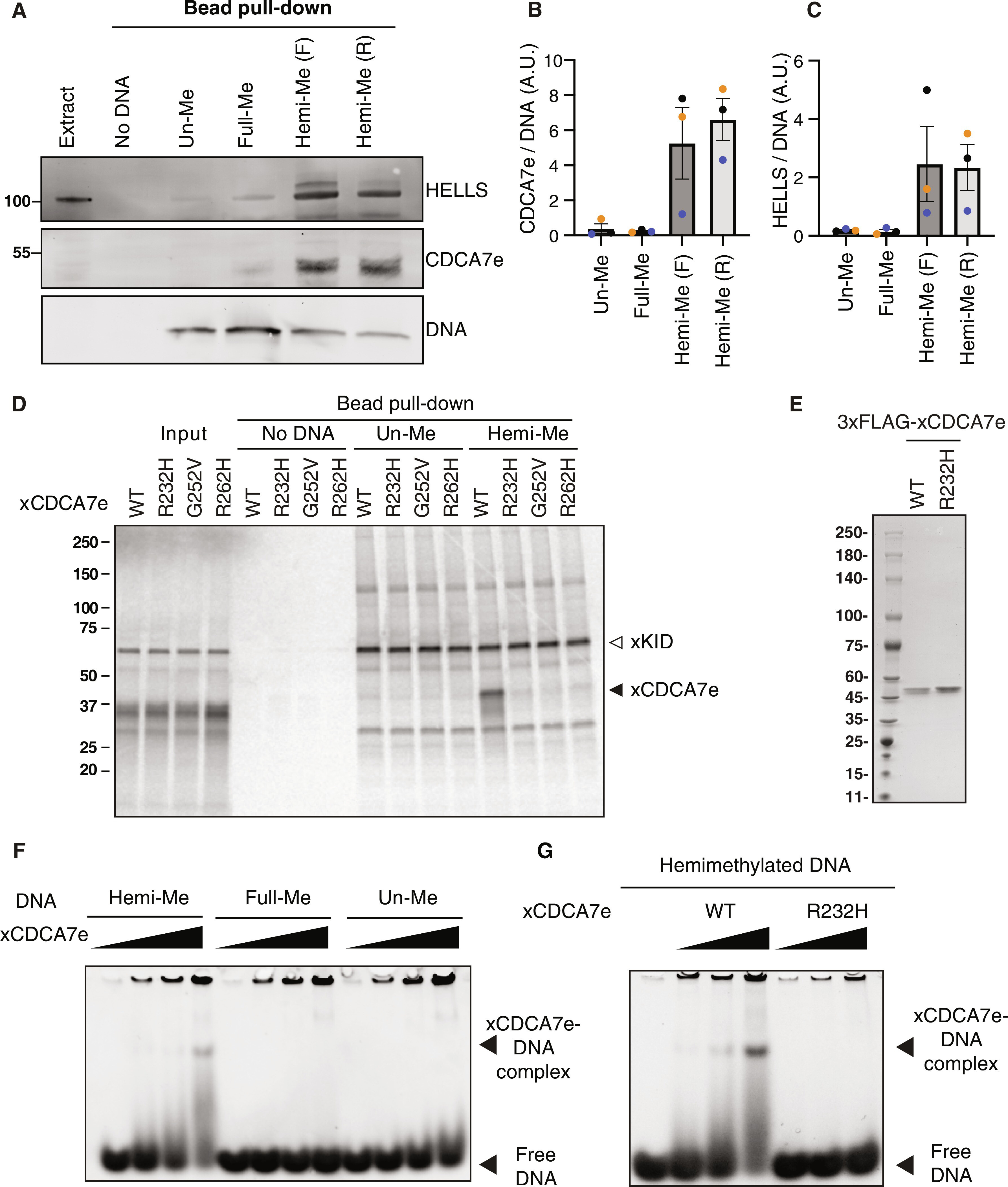
Fig. 1. CDCA7 selectively binds hemimethylated DNA.
(A) Magnetic beads coupled with double-stranded 54-bp DNA oligos containing unmethylated CpGs (un-Me), fully methylated CpGs (full-Me), or hemimethylated CpGs [hemi-Me; (F) and (R) to indicated 5mC in the forward- or reverse- strand; table S1], were incubated with interphase Xenopus egg extracts. Beads were collected after 10 min and analyzed by Western blotting. SDS–polyacrylamide gel electrophoresis (SDS-PAGE) was stained with SYBR Safe to visualize loading of the 54-bp DNA. Representative of n = 3 independent experiments. (B) Quantification of CDCA7e signal in Western blot analyses described in (A). CDCA7e signal at the DNA beads is normalized relative to the DNA signal. A.U., arbitrary units. n = 3 (biological replicates). The means and SEM are shown. (C) Quantification of HELLS signal in the Western blot analyses described in (A). HELLS signal at the DNA beads is normalized relative to the DNA signal. n = 3 (biological replicates). The means and SEM are shown. (D) 35S-labeled X. laevis CDCA7e proteins (wild type or with the indicated ICF3-patient associated mutation) were incubated with control beads, or beads conjugated 200-bp unmethylated or hemimethylated DNA (table S1). 35S-labeled xKid (80), a nonspecific DNA binding protein, was used as a loading control. Autoradiography of 35S-labeled proteins in input and beads fraction is shown. (E) Coomassie staining of purified 3xFLAG-tagged CDCA7eWT and CDCA7eR232H used in (F) and (G). (F and G) EMSA using recombinant X. laevis (F) CDCA7eWT and (G) CDCA7eR232H. In graphs, data points from each biological replicate are annotated in a unique color.

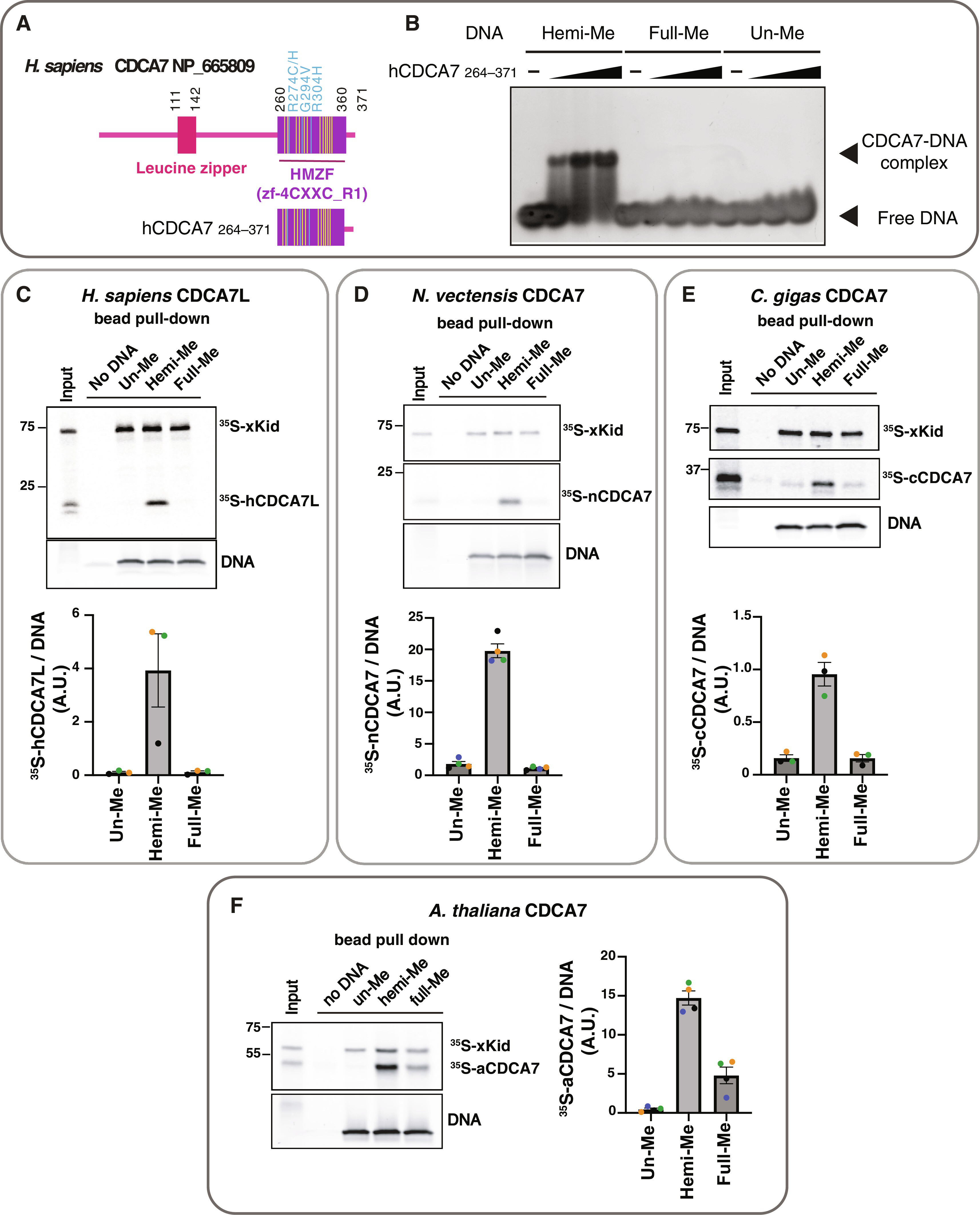
Fig. 2. Selective binding of hemimethylated CpG by the HMZF domain of CDCA7 is evolutionarily conserved.
(A) Schematic of H. sapiens CDCA7 (isoform 2 NP_665809). Positions of the HMZF (zf-4CXXC_R1) domain (purple), three ICF3-patient mutations (cyan), and conserved cysteine residues (yellow) are shown. (B) EMSA assay using the purified HMZF domain (amino acids 264 to 371) of H. sapiens CDCA7. (C to F) Magnetic beads coupled with double-stranded 54-bp DNA oligos containing unmethylated (un-Me), hemimethylated (hemi-me), or fully methylated (full-Me) CpGs were incubated for 10 min in the presence of the indicated 35S-labeled CDCA7 homolog and 35S-labeled xKid proteins. SDS-PAGE gels were stained with SYBR-Safe to visualize loading of the 54-bp DNA. Representative autoradiographs of 35S-labeled proteins in input and DNA pull-downs are shown. Quantifications of pulled-down 35S CDCA7 signal relative to the DNA signal are shown in a bar graph indicating the mean with SEM. In graphs, data points from each biological replicate are annotated in a unique color. The means and SEM are shown. (C) Pull-down of 35S-labeled human CDCA7 paralog CDCA7L (amino acids 322 to 454 of NP_061189) from Xenopus egg extract. Bar graph shows the quantification of data from n = 3 independent experiments. (D) Pull-down of 35S-labeled N. vectensis CDCA7 homolog (EDO33918.1) from boiled and clarified Xenopus egg extract supernatant. Bar graph shows the quantification of data from n = 4 independent experiments. (E) Pull-down of 35S-labeled C. gigas CDCA7 homolog (XP_011438013) from boiled and clarified Xenopus egg extract supernatant. Bar graph shows quantification of data from n = 3 independent experiments. (F) Pull-down of 35S-labeled A. thaliana CDCA7 homolog (NP_195428) from boiled and clarified Xenopus egg extract supernatant. Bar graph shows quantification of data from n = 4 independent experiments.

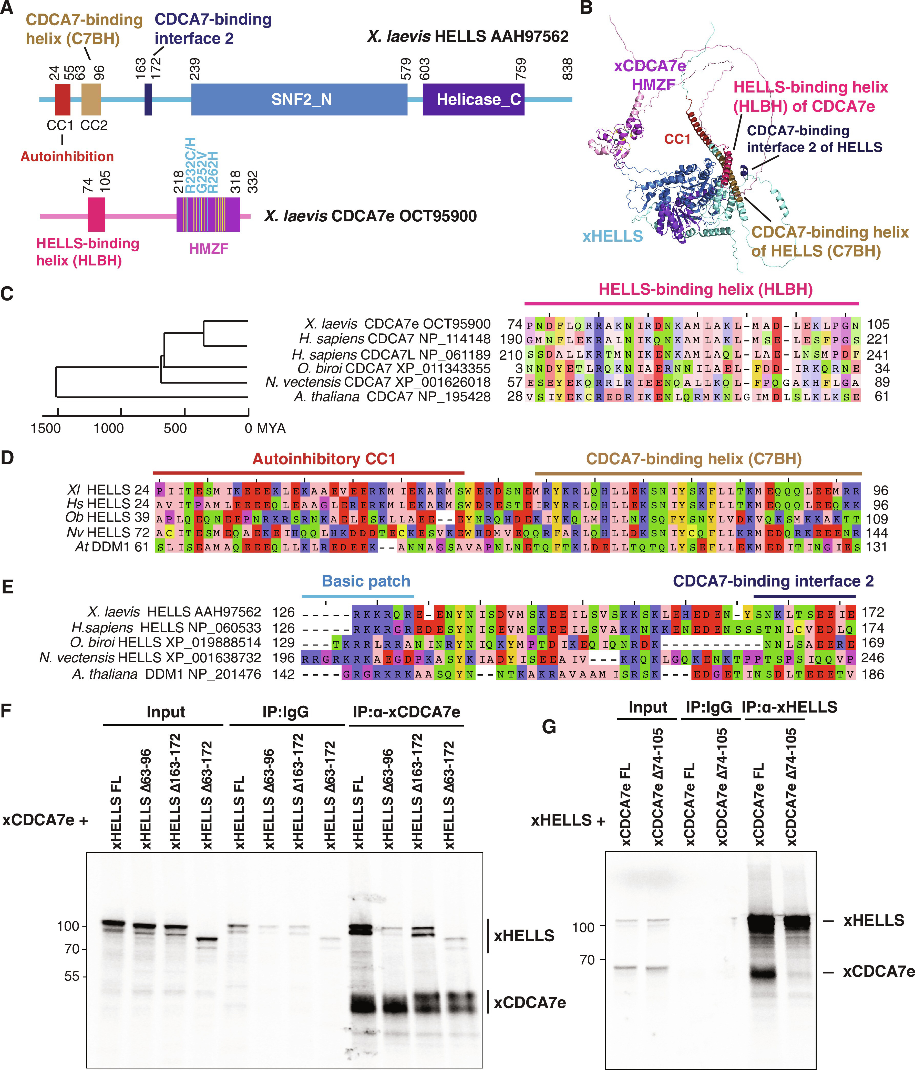
Fig. 5. Identification of HELLS-CDCA7 interaction interface.
(A) Schematics of X. laevis HELLS and CDCA7e. Positions of the signature 11 conserved cysteine residues and 3 ICF disease–associated mutations in CDCA7e are marked in yellow and cyan, respectively. CC1 is a coiled-coil domain important for autoinhibition. (B) The best predicted structure model of X. laevis HELLS-CDCA7e complex by AF2. (C) Sequence alignment of the putative HELLS/DDM1-binding interface of CDCA7. (D) Sequence alignment of the putative CDCA7-binding interface 1 in HELLS/DDM1. (E) Sequence alignment of the putative CDCA7-binding interface 2 in HELLS. (F) Immunoprecipitation by control IgG or anti-CDCA7e antibodies from Xenopus egg extracts containing 35S-labeled wild-type or deletion mutant of X. laevis HELLS and CDCA7e. (G) Immunoprecipitation by control IgG or anti-HELLS antibody from Xenopus egg extracts containing 35S-labeled HELLS and wild-type or ∆74-105 deletion mutant of CDCA7e. Autoradiography is shown in (F) and (G).
Adapted with permission from AAAS on behalf of Science Advances: Wassing et al. (2024). CDCA7 is an evolutionarily conserved hemimethylated DNA sensor in eukaryotes. Sci Adv 2024 Aug 23;1034:eadp5753. doi: 10.1126/sciadv.adp5753.
This work is licensed under a Creative Commons Attribution 4.0 International License. The images or other third party material in this article are included in the article’s Creative Commons license, unless indicated otherwise in the credit line; if the material is not included under the Creative Commons license, users will need to obtain permission from the license holder to reproduce the material. To view a copy of this license, visit http://creativecommons.org/licenses/by/4.0/
Last Updated: 2024-10-15