Colorless and immunodeficient frogs - a valuable platform for tumor and developmental biology research
Revealing mitf functions and visualizing allografted tumor metastasis in colorless and immunodeficient Xenopus tropicalis
Commun Biol 2024 Mar 05;71:275. doi: 10.1038/s42003-024-05967-3.
Ran R, Li L, Xu T, Huang J, He H, Chen Y.
Click here to view article at Communications Biology.
Click here to view article at Pubmed.
Click here to view article on Xenbase.
Abstract
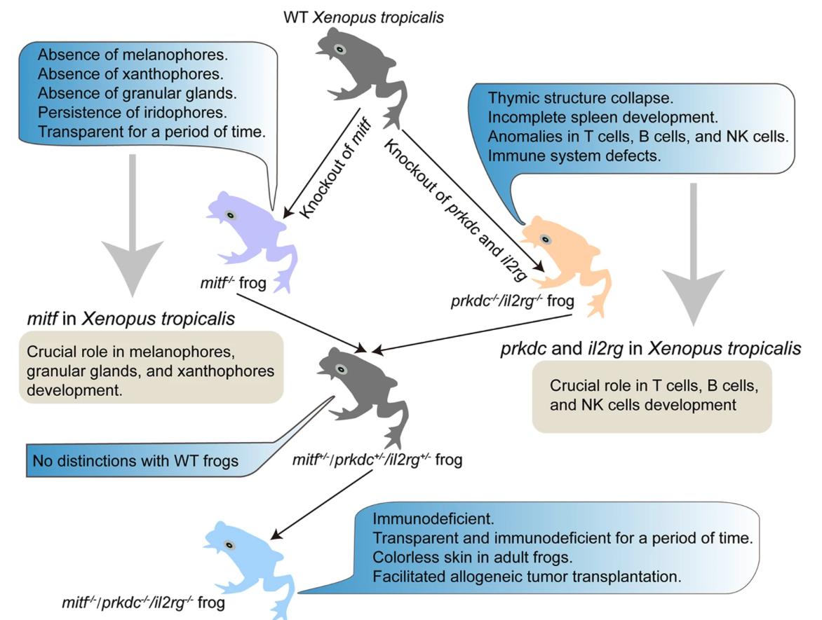
Transparent immunodeficient animal models not only enhance in vivo imaging investigations of visceral organ development but also facilitate in vivo tracking of transplanted tumor cells. However, at present, transparent and immunodeficient animal models are confined to zebrafish, presenting substantial challenges for real-time, in vivo imaging studies addressing specific biological inquiries. Here, we employed a mitf-/-/prkdc-/-/il2rg-/- triple-knockout strategy to establish a colorless and immunodeficient amphibian model of Xenopus tropicalis. By disrupting the mitf gene, we observed the loss of melanophores, xanthophores, and granular glands in Xenopus tropicalis. Through the endogenous mitf promoter to drive BRAFV600E expression, we confirmed mitf expression in melanophores, xanthophores and granular glands. Moreover, the reconstruction of the disrupted site effectively reinstated melanophores, xanthophores, and granular glands, further highlighting the crucial role of mitf as a regulator in their development. By crossing mitf-/- frogs with prkdc-/-/il2rg-/- frogs, we generated a mitf-/-/prkdc-/-/il2rg-/- Xenopus tropicalis line, providing a colorless and immunodeficient amphibian model. Utilizing this model, we successfully observed intravital metastases of allotransplanted xanthophoromas and migrations of allotransplanted melanomas. Overall, colorless and immunodeficient Xenopus tropicalis holds great promise as a valuable platform for tumorous and developmental biology research.
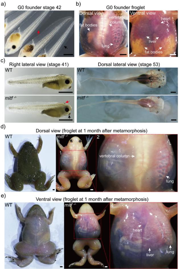
Fig. 1: Biallelic mitf disruption makes Xenopus tropicalis transparent within two months post-metamorphosis. a After using the CRISPR/Cas9 system to knock out mitf via gRNA T7, founder generation (G0) mosaic tadpoles at stage 42 exhibited a loss of melanophores (indicated by red arrow), compared to wild type (WT) tadpoles from the same batch (indicated by black arrow). 79 mosaic tadpoles and 20 wild type tadpoles were observed. b One month after metamorphosis, the transparent skin of the mosaic froglet allowed for external visibility of internal organs such as the fat bodies, lung, liver, heart, and vertebral column. Here was shown one representative froglet, out of a total of 11 mosaic froglets. c The F1 mitf−/− Xenopus tropicalis tadpole exhibited melanophores loss throughout the entire body at stages 41 and 53 compared to wild type tadpoles from the same batch. Notably, at stage 41, the melanin pigmentation in the eyes of mitf−/− Xenopus tropicalis was generated by retinal pigment epithelium (RPE) cells, and redistribution of melanin in oocytes resulted in some melanin pigment in the head (indicated by red arrow) and cement gland (indicated by black arrow). However, the melanin pigment in the head and cement gland disappeared in later development stages, such as stage 53. Representative photographs were exhibited from 35 F1 mitf−/− Xenopus tropicalis tadpoles at stage 41, 15 F1 mitf−/− Xenopus tropicalis tadpoles at stage 53, as well as from 10 wild type tadpoles each at stage 41 and 53. d, e One month after metamorphosis, the F1 mitf−/− Xenopus tropicalis froglet exhibited transparent skin from both dorsal (d) and ventral (e) views. Red dashed boxes 2 and 4 corresponded to magnified views of red dashed boxes 1 and 3, respectively. The transparent skin allowed for external visibility of internal organs including the lung, liver, heart, and vertebral column within one month after metamorphosis as shown. Ten F1 mitf−/− Xenopus tropicalis froglets and ten wild-type Xenopus tropicalis froglets were used to provide representative photographs as shown. Each scale bar is 1 mm.
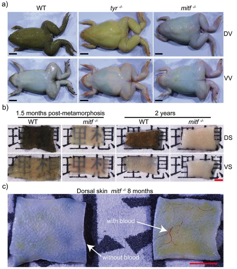
Fig. 2: The skin of mitf−/− adult frogs turns colorless and opaque. a The representative photographs of adult wild type (WT), tyr−/−, and mitf−/− Xenopus tropicalis were displayed to showcase their dorsal view (DV) and ventral view (VV). The photographs were obtained from a sample of 15 adult frogs of each genotype, all of which were 1 year old. b Skin samples approximately 2 mm×5 mm in size were obtained from both dorsal skin (DS) and ventral skin (VS) of wild-type and mitf−/− Xenopus tropicalis aged 2 years or 1.5 months postmetamorphosis. These samples were utilized in transillumination experiments to assess their translucency properties. Specifically, the experiment involved positioning each sample on a sheet of paper with the word “理想“ and photographing them under consistent lighting and camera settings to assess the visibility of the word. Three frogs were used for each genotype, and one sample of DS and VS skin was collected per frog. Representative photographs of each genotype were presented to illustrate the experimental findings. c Skin samples were obtained with blood or after exlusion of blood from dorsal regions of the same mitf−/− Xenopus tropicalis aged 8 months. Three frogs were used for this experiment. The representative photograph was presented. The black scale bar in a is 1 cm. The red scale bar in b and c is 1 mm.
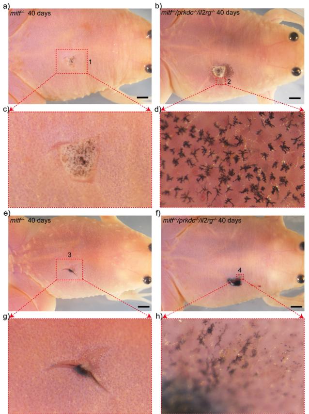
Fig. 10: Allogeneic transplantations of melanomas were conducted in mitf−/−/prkdc−/−/il2rg−/− Xenopus tropicalis, resulting in the observed migrations of transplanted melanomas in the recipient animals. a–h At day 40 post-transplantation, the transplanted nevi and metastatic melanoma were examined in mitf−/− (a, c, e, g) and mitf−/−/prkdc−/−/il2rg−/− (b, d, f, h) recipients. The enlarged views of the transplanted nevi and metastatic melanoma were portrayed in panels c and d (red dashed boxes 1 and 2) and g and h (red dashed boxes 3 and 4), respectively. Each scale bar is 1 mm.
Adapted with permission from Communications Biology on behalf of Springer Nature: Ran et al. (2024). Revealing mitf functions and visualizing allografted tumor metastasis in colorless and immunodeficient Xenopus tropicalis.Commun Biol 2024 Mar 05;71:275. doi: 10.1038/s42003-024-05967-3.
This work is licensed under a Creative Commons Attribution 4.0 International License. The images or other third party material in this article are included in the article’s Creative Commons license, unless indicated otherwise in the credit line; if the material is not included under the Creative Commons license, users will need to obtain permission from the license holder to reproduce the material. To view a copy of this license, visit http://creativecommons.org/licenses/by/4.0/
Last Updated: 2024-08-22