Discovering Frog Development in Unprecedented Detail
Unveiling vertebrate development dynamics in frog Xenopus laevis using micro-CT imaging
Gigascience 2024 Jan 02;13. doi: 10.1093/gigascience/giae037.
Laznovsky J, Kavkova M, Helena Reis A, Robovska-Havelkova P, Maia LA, Krivanek J, Zikmund T, Kaiser J, Buchtova M, Harnos J.
Click here to view article at Gigascience.
Click here to view article at PubMed.
Click here to view article on Xenbase.
Interesting write-up in Phys.org: New 3D anatomical atlas of the African clawed frog increases understanding of development and metamorphosis processes
Graphical Abstract
Summary: X-ray tomography was used to examine the African clawed frog (Xenopus laevis). This extensive dataset of specimens from tadpoles to adult frogs opens avenues to novel insights into the changes and developmental dynamics of selected structures, leading eventually to an improved understanding of this crucial animal model.
Laznovsky et al. used advanced micro-CT imaging to explore the developmental stages of the African clawed frog, Xenopus laevis. By generating high-resolution 3D models from tadpoles to adult frogs, they created a comprehensive dataset enhancing our understanding of vertebrate development. This resource offers valuable insights into the morphological changes during frog development and is suitable for various research applications, educational purposes, and even virtual reality use.
A 3D atlas of the African Clawed Frog
High-quality X-ray microtomography images cover the lifespan of the African Clawed Frog in unprecedented detail.
A 3D anatomical atlas of the model organism Xenopus laevis (the African clawed frog) helps researchers to better understand embryonic development, including metamorphosis – the intriguing process by which a tadpole transforms into a mature frog. The anatomical atlas, published in the open science journal GigaScience, has also been converted into a set of 3D printable models that are available from the digital design platform Thingiverse.
From transplantation experiments in the early twentieth century up to the present age of genome sequencing, the African Clawed Frog (Xenopus laevis) is a versatile vertebrate model organism for developmental biology and other disciplines. It is particularly suited to study body plan reorganization during the big changes that happen when the tadpole transforms into a mature frog (“metamorphosis”). “However, a notable gap exists in the availability of comprehensive datasets encompassing Xenopus’ late developmental stages”, explains Dr. Jakub Harnos from Masaryk University (Czech Republic), a leading scientist of the GigaScience study.
To add this missing data and to more accurately describe the multiple stages of Xenopus laevis development, the authors used X-ray microtomography, a 3D imaging technique, to create an anatomical atlas. Through detailed analysis of the 3D reconstructions at the various stages of development, the authors could pinpoint key changes that occur during the developmental journey from tadpole through to froglet and mature adult stages.
One striking example of the shape changes that can be tracked in great detail with this new high-resolution data is the adjustment of the position of the developing frog’s eyes, and the exact timing of this shift. With advancing development, the distance between the eyes progressively decreases. “This adaptation aligns well with the frog's life strategy, transitioning from a water-dwelling tadpole with lateral eyes to an adult with eyes positioned on top of the head for a submerged lifestyle, reminiscent of crocodilians”, the authors note.
Also, the frog’s gut undergoes significant remodeling during metamorphosis. Over an 8-day period, the intestine shortens by around 75 %, and the coiling pattern changes drastically – a process that can be followed in fine detail by X-ray microtomography but would be hard to study with other methods. Other anatomical facts that are revealed in great detail by the new 3D atlas concern the differences between male and female frogs (females end up larger, overall) and the very subtle positioning of the teeth of Xenopus laevis, which are hidden behind the maxillary arch.
Nevertheless, the main advantage of this study is that it provides all X-ray data openly, allowing other (Xenopus) researchers to investigate more both soft and hard tissues in unprecedented detail in this key vertebrate model.
To enable other researchers, and also the 3D printing community, to easily access printable models, a collection of 40 surface-rendered 3D models from the Xenopus laevis anatomical atlas is available from the design platform Thingiverse.
Printable 3D models are additionally available from SketchFab and Thingiverse.
The SketchFab collections:
Xenopus development: https://sketchfab.com/GigaDB/collections/xenopus-laevis-development-3922d82fde27407ea1e7cc4622376178.
Xenopus adult male: https://sketchfab.com/GigaDB/collections/xenopus-laevis-male-7eb069e65d2f49749ce4b4144dd5fc81.
Xenopus adult female: https://sketchfab.com/GigaDB/collections/xenopus-laevis-female-b33299ca19604663a0cbdeb915f683e9.
The Thingiverse links for the collection of printable models: https://www.thingiverse.com/thing:6620040.
Supporting data for "Unveiling Vertebrate Development Dynamics in Frog Xenopus laevis using Micro-CT Imaging"
The GigaDB repository provide efficient accessibility through 8-bit TIFF stacks, complemented by .STL models, with recommended 3D explorationusing software like VGStudioMAX, MyVGL ,Avizo, or Fiji ImageJ.
Data stored at the GigaDB repository: http://gigadb.org/dataset/102534.
Data stored at Zenodo: https://zenodo.org/records/10951411.
Data stored on Xenbase FTP: https://bigfrog2.xenbase.org/pub/xenbase/images/Laznovsky_3D_Atlas/.
The dataset contains X-ray Micro Computed Tomography data of Xenopus laevis frog. There are twenty datasets of ten individual animals.
Each animal was CT scanned twice – once as a native scan to visualize the hard tissues, and once contrast-stained to visualize the soft tissues.
The datasets include nine developmental stages (NF44-45, NF52, NF53, NF54, NF57, NF59, NF62, NF66 and adult).
There are two adults, one male and one female.
The CT data (in 8bit .tiff format compressed as .tar.gz files) are supported by .stl files created from each dataset.
The database also includes .stl files of selected structures of interest (body, skeleton, skull, brain and guts of individual animals).
Abstract
BACKGROUND: Xenopus laevis, the African clawed frog, is a versatile vertebrate model organism in various biological disciplines, prominently in developmental biology to study body plan reorganization during metamorphosis. However, a notable gap exists in the availability of comprehensive datasets encompassing Xenopus' late developmental stages. FINDINGS: This study utilized micro-computed tomography (micro-CT), a noninvasive 3-dimensional (3D) imaging technique with micrometer-scale resolution, to explore the developmental dynamics and morphological changes in Xenopus laevis. Our approach involved generating high-resolution images and computed 3D models of developing Xenopus specimens, spanning from premetamorphosis tadpoles to fully mature adults. This dataset enhances our understanding of vertebrate development and supports various analyses. We conducted a careful examination, analyzing body size, shape, and morphological features, focusing on skeletogenesis, teeth, and organs like the brain and gut at different stages. Our analysis yielded valuable insights into 3D morphological changes during Xenopus' development, documenting details previously unrecorded. These datasets hold the solid potential for further morphological and morphometric analyses, including segmentation of hard and soft tissues. CONCLUSIONS: Our repository of micro-CT scans represents a significant resource that can enhance our understanding of Xenopus' development and the associated morphological changes in the future. The widespread utility of this amphibian species, coupled with the exceptional quality of our scans, which encompass a comprehensive series of developmental stages, opens up extensive opportunities for their broader research application. Moreover, these scans can be used in virtual reality, 3D printing, and educational contexts, further expanding their value and impact.
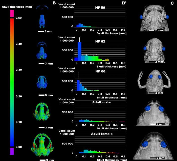
Figure 2
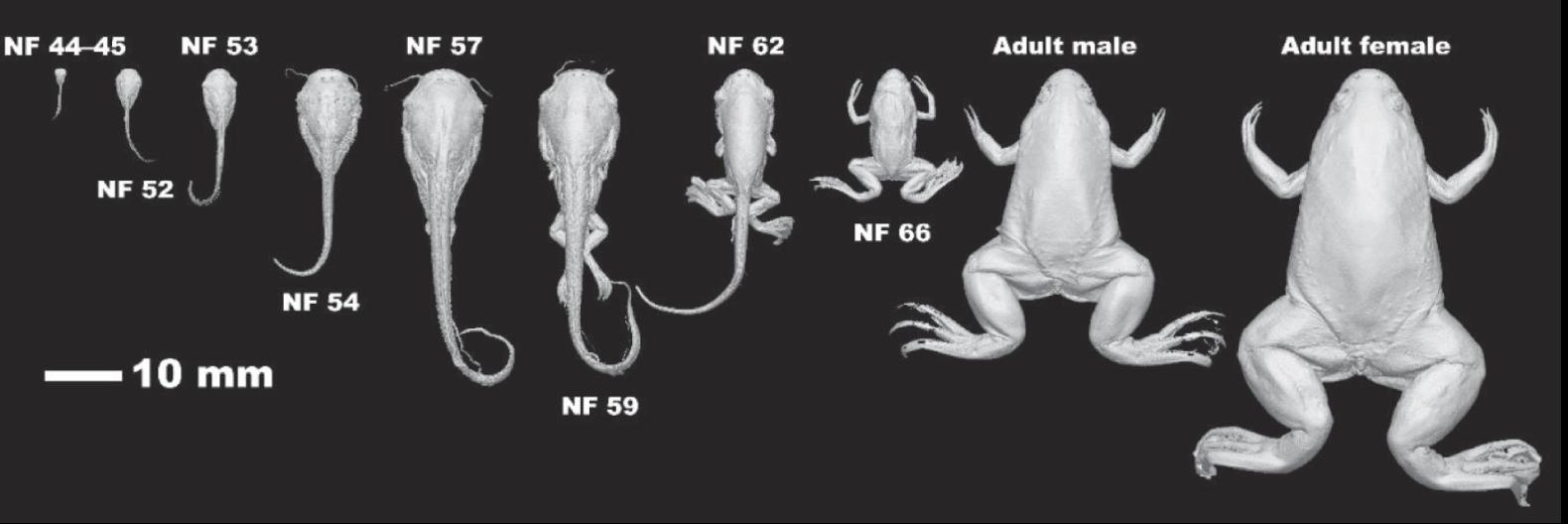
The analyses of the head development. (A) The series of Xenopus skulls of selected stages. The scale bar with values in mm is shown on the left bottom. (B) The skull thickness of selected stages is displayed. The scale bar with values in mm is shown on the left side. (B′) The relative distribution of bone thickness for each skull is shown. The experiment involved conducting 3 repetitions for each developmental stage, with the results depicted as means accompanied by standard deviations (± SD). (C) The overview of developing heads with highlighted eyes is shown.
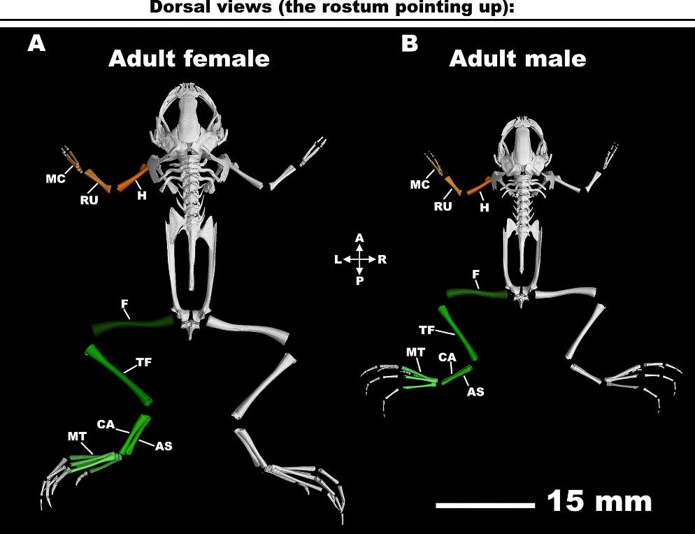
Figure 5
The analysis of frog skeletogenesis. (A) The adult male frog with analyzed bones is depicted. (B) The adult female frog with analyzed bones is displayed. (C) The graph demonstrating the analyzed bones throughout the Xenopus laevis’ late development. The experiment involved conducting 3 repetitions for each long bone measurement, with the results depicted as means accompanied by standard deviations (± SD). However, error bars are depicted only for cases demonstrating statistically significant differences, while they are not displayed for insignificant variations. Statistical analysis was performed using a 2-way analysis of variance (ANOVA) followed by Tukey post hoc test for multiple comparisons; ****P < 0.0001. A: anterior; AS: astragalus; CA: calcaneum; F: femur; H: humerus; L: left; MC: metacarpals; MT: metatarsals; P: posterior; R: right; RU: radioulna; TF: tibiofibular.
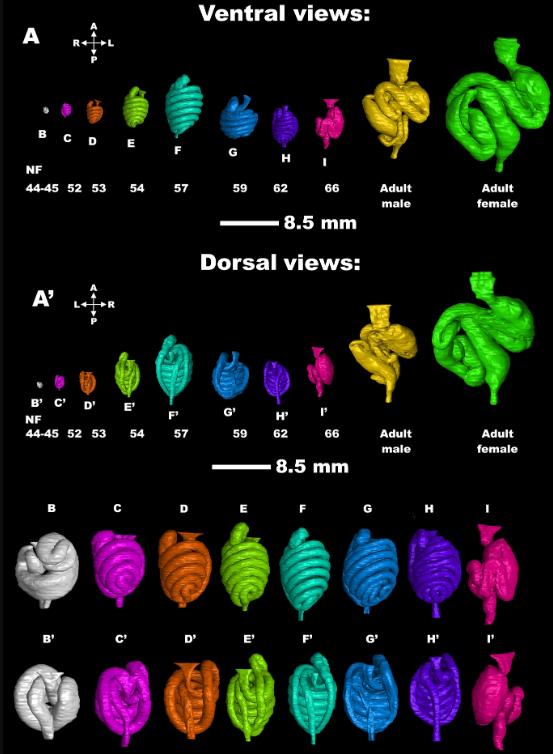
Figure 8
The analysis of gut development. (A–A′) Ventral and dorsal views of individual stages of Xenopus gut and their details for each developmental stage (B–I, B′–I′), except guts from frog adults.
Adapted with permission from Oxford University Press on behalf of Gigascience: Laznovsky et al. (2024). Unveiling vertebrate development dynamics in frog Xenopus laevis using micro-CT imaging.Gigascience 2024 Jan 02;13. doi: 10.1093/gigascience/giae037.
This work is licensed under a Creative Commons Attribution 4.0 International License. The images or other third party material in this article are included in the article’s Creative Commons license, unless indicated otherwise in the credit line; if the material is not included under the Creative Commons license, users will need to obtain permission from the license holder to reproduce the material. To view a copy of this license, visit http://creativecommons.org/licenses/by/4.0/
Last Updated: 2024-07-22