XB-IMG-46625
Xenbase Image ID: 46625

|
||||||||||
|
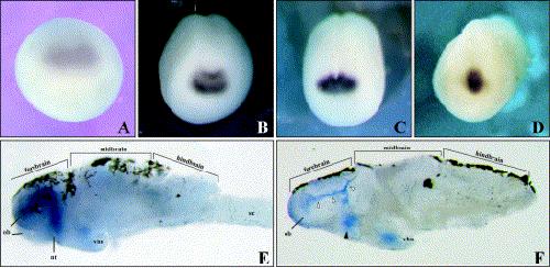
Fig. 2. Fez expression pattern in Xenopus embryos and tadpole brain. (AâD) Anterior views (top is dorsal) of embryos at stage 12 (A), stage 13/14 (B), stage 16/17 (C), and stage 24 (D). The signal intensifies during development and remains localized to the telencephalic region. (E) A lateral view of Xenopus brain at stage 45, showing the approximate boundaries of the forebrain, midbrain, and hindbrain. A strong expression is detected in the forebrain. (F) A saggital section of the Xenopus brain at the same stage, displaying expression in the olfactory bulbs, nervus terminalis (black arrowhead) and ventricular zone (white arrowheads). A faint expression is visible near the ventral hypothalamic nucleus. Brown fiber-like processes at the top of the brain in (E,F) are epidermal pigments. Abbreviations: nt, nervus terminalis; ob, olfactory bulb; sp, spinal cord; vhn, ventral hypothalamic nucleus. Image published in: Matsuo-Takasaki M et al. (2000) Copyright © 2000. Image reproduced with permission of the Publisher, Elsevier B. V.
Image source: Published Larger Image Printer Friendly View |
