XB-IMG-45878
Xenbase Image ID: 45878

|
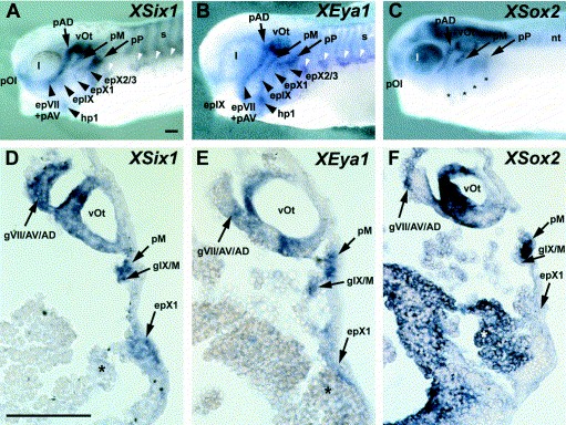
Fig. 4. Expression of XEya1 (B,E) compared to other placodal markers XSix1 (A,D) and XSox2 (C,F) in lateral views (AâC) and transverse sections at the level of the otic vesicle (DâF) of stage 30â34 Xenopus embryos. Patterns of expression of XEya1 (B,E) and XSix1 (A,D) are largely identical in the adenohypophysis (data not shown), olfactory placodes (pOl), the otic vesicle(vOt), lateral line placodes (pAV, pAD, pM, pP), epibranchial (epVII, epIX, epX1, epX2/3) and hypobranchial placodes (hp1). Co-expression of both genes is also observed in cranial ganglia that have a placodally-derived component, e.g. in the profundal/trigeminal ganglionic complex (data not shown), in the fused ganglia of the facial, anteroventral and anterodorsal lateral line nerves (gVII/AV/AD in D,E), and in the fused ganglia of the glossopharyngeal and middle lateral line nerves (gIX/M in D,E). Additionally, XEya1 and XSix1 are co-expressed in the somites (s in A,B), in hypaxial muscle precursors (white arrowheads in A,B) and weakly in the pharyngeal pouches (asterisks in D,E). Placodal expression of XSox2 (C,F) overlaps with the expression of XEya1 and XSix1 in the adenohypophysis (data not shown), the olfactory placode, the otic vesicle and the lateral line placodes, as well as in some cranial ganglia (e.g. gVII/AV/AD in F). In contrast to XEya1 and XSix1, however, XSox2 is not expressed in the profundal/trigeminal placodes or ganglia (data not shown) and in the epibranchial placodes (C), whereas it is strongly expressed in the neural tube (nt), the lens (l), and the pharyngeal pouches (asterisks). Bar in (A): 0.1 mm (AâC). Bar in (D): 0.1 mm (DâF). Image published in: David R et al. (2001) Copyright © 2001. Image reproduced with permission of the Publisher, Elsevier B. V.
Image source: Published Larger Image Printer Friendly View |
