XB-IMG-45411
Xenbase Image ID: 45411

|
|||||||||||||||||||||||||||||||||||||||||||||
|
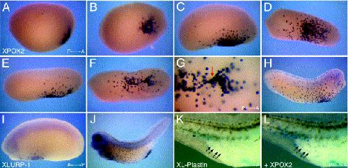
Fig. 2. Whole-mount in situ hybridisation analysis of Xenopus POX2 (AâH,L), LURP-1 (I,J) and L-plastin (K) expression. Right-lateral views of stage 19 (A), 24 (C) and 27 (E) embryos showing XPOX2-expressing cells. (B,D,F) Ventral views of the same embryos depicted in (A,C,E). (G) Higher magnification image of the stage 27 embryo depicted in (F) to illustrate the streams of XPOX2-expressing cells that emanate from a focal point (red arrow) located at the ventral midline within the heart-forming region. (H) Right-lateral view of a stage 30 embryo. (I,J) Left-lateral views of embryos at stages 24 and 36, respectively, showing cells that express XLURP-1. (K,L) Right-lateral view of the posterior trunk of a stage 35 tadpole after sequential, double whole-mount in situ hybridisation staining to reveal first (K), Xenopus L-plastin (pale blue) and second (L), POX2 (magenta) expression. The co-localisation of the two chromogenic reagents results in a dark blue colour (L). A, anterior; P, posterior. Image published in: Smith SJ et al. (2002) Copyright © 2002. Image reproduced with permission of the Publisher, Elsevier B. V.
Image source: Published Larger Image Printer Friendly View |
