XB-IMG-3322
Xenbase Image ID: 3322
|
||||||||||
|
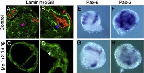
Fig. 5. Depletion of Dg affects laminin-1 assembly and tubule formation in vitro. Two-cell stage embryos were injected with Dg-Mo1Â +Â 2 (32 ng/embryo). Animal caps were treated with activin/retinoic acid for 3 h. (A, D) Immunodetection with the 3G8 antibodies and the anti-laminin antibodies. (A, B) Section of a control animal cap. 3G8 (blue arrowhead) and anti-laminin-1 (red arrowhead) antibodies staining are detected. Laminin-1 is present at the basal pole of cells of the tubules. Laminin-1 is also observed elsewhere in the explant where the tubules will be formed (purple arrowhead) (A). (C, D) Section of a Mo1Â +Â 2 injected explant (C). Large cavities and mesenchyme like tissues are observed. (D) A diffuse fluorescence is observed (white arrowhead) suggesting that laminin-1 is accumulated but does not assemble basal lamina. (EâH) Expression of pronephros marker genes in animal caps. (E, G) The expression of the Pax-8 gene is similar in the control and Mo1Â +Â 2 injected explant. (F, H) Pax-2 gene is detected in control and Mo1Â +Â 2 injected explant. Image published in: Bello V et al. (2008) Copyright © 2008. Image reproduced with permission of the Publisher, Elsevier B. V.
Image source: Published Larger Image Printer Friendly View |