XB-IMG-192517
Xenbase Image ID: 192517
|
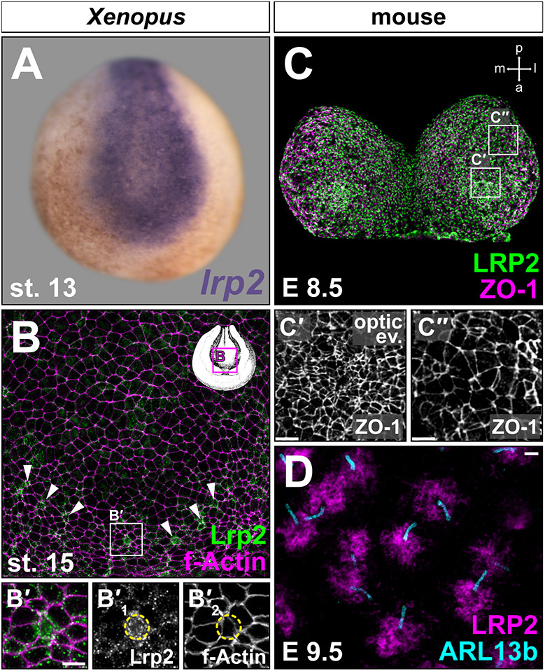
Fig. 1.
Lrp2 is expressed in the neuroepithelium and increased in constricting cells. (A-D) lrp2 mRNA (A) and protein (B-D) expression analyzed by in situ hybridization and immunofluorescence, respectively. Neurula stage (st.) embryos (frontal views, dorsal upwards). (A) Neural lrp2 expression. (B) Stage 15 forebrain region; Lrp2 is expressed in most cells (outlined by F-actin); single cells with high Lrp2 levels are located along the anterior rim of neural folds (NFs; arrowheads). (Bâ²) Magnification of boxed region in B; increased Lrp2 levels are found in cells with small apical surface (circled in single channels Bâ²1 and Bâ²2). (C) LRP2 is detected throughout E8.5 anterior NFs, concentrated in areas undergoing apical constriction. Compare Câ² (constricted cells in optic evagination) with Câ³ (dorsolateral cells with larger cell surface). ZO-1 marks cell boundaries. (D) STED imaging; LRP2 is condensed around neuroepithelial primary cilia (ARL13b+) at E9.5. Scale bars: 10â
µm in Bâ²,Câ²,Câ³; 1â
µm in D. Image published in: Kowalczyk I et al. (2021) Copyright © 2021. Image reproduced with permission of the Publisher and the copyright holder. This is an Open Access article distributed under the terms of the Creative Commons Attribution License.
Image source: Published Larger Image Printer Friendly View |