XB-IMG-174600
Xenbase Image ID: 174600
|
|||||||||||||||
|
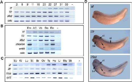
Fig. 2. Temporal and spatial expression ofXenopus laevis lifand lif receptors encoded genes. (A) RT-PCR analysis of embryo from stage 2 to stage 33. (B) RT-PCR analysis of dissected parts of stage 11 embryo. AC, animal cap; Do, dorsal mesoderm; Em, total embryo; En, endoderm; Ve, ventral mesoderm. Chordin and wnt8 expression is used as control. (C) RT-PCR analysis of adult tissues. Br, brain; He, heart; Ki, kidney; Li, liver; Lu, lung; Mu, skeletal muscle; Ov, ovary; Sk, skin; Sp, spleen; St, stomach; Te, testis. Ornithine decarboxylase (odc) and ribosomal protein L8 (rpl8) gene expression was used as control. - Control without reverse transcription. (D) In situ hybridization on stage 37/38 embryo with lif, lifr and il6st antisense probes. Ba, branchial arches; ki, embryonic kidney; nt, neural tube; ot, otic vesicle; so, somites. Image published in: Jalvy S et al. (2019) Copyright © 2019. Image reproduced with permission of the Publisher, Elsevier B. V.
Image source: Published Larger Image Printer Friendly View |