XB-IMG-135816
Xenbase Image ID: 135816
|
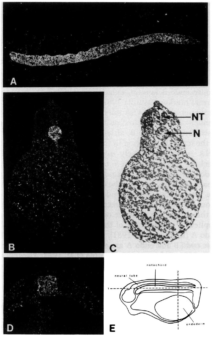
Figure 4. In situ hybridization to sections of Xenopus embryos.
[A] Dark-field illumination shows hybridization of
NC-11 throughout the notochord from the anterior to posterior
region of a stage 25 embryo (see £). [B] Dark-field illumination
shows hybridization to the notochord in a stage 25 embryo. (C)
Bright-field illumination of the section shown in B. (N) Notochord;
(NT) neural tube. (D) Dark-field illumination of a stage
14 embryo shows hybridization to the notochord. (£) Plane of
sections shown in A and B. Although the embryos are at different
stages, the plane of section in D is the same as that in B. Image published in: LaFlamme SE et al. (1988) Copyright © 1988. Image reproduced on with permission of the Publisher, Cold Spring Harbor Laboratory Press. This is an Open Access article.
Image source: Published Larger Image Printer Friendly View |