Click here to close Hello! We notice that you are using Internet Explorer, which is not supported by Xenbase and may cause the site to display incorrectly. We suggest using a current version of Chrome, FireFox, or Safari.
XB-IMG-86862

Xenbase Image ID: 86862


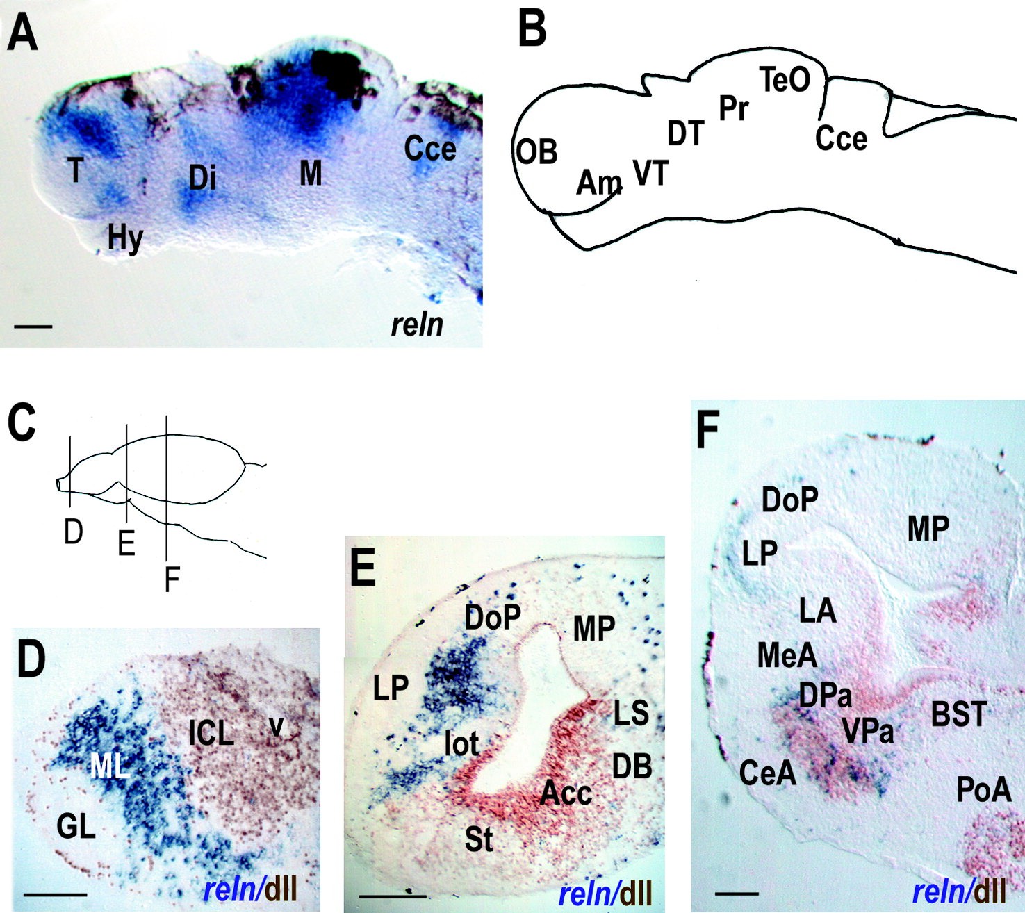
Figure 9. Reelin expression in the brain of developing Xenopus laevis. A,B:reelin (reln) expression (A) and schematic drawing (B) of stage 35 Xenopus embryo. C: Schematic drawing of a stage 50 Xenopus brain, illustrating the position of sections in D–F. D–F: Frozen transverse sections showing gene and/or protein expression (indicated bottom right). Dorsal is to the top. D: Accessory olfactory bulb. E: Rostral telencephalon. F: Caudal telencephalon. For abbreviations , see list. Scale bars = 100 μm in A, D–F.

Image published in: Costagli A et al. (2002)

Copyright © 2002. Image reproduced with permission of the Publisher, John Wiley & Sons.

GeneSynonymsSpeciesStage(s)Tissue
reln.LreelinX. laevisThroughout NF stage 35 and 36brain
telencephalon
diencephalon
midbrain
optic tectum
cerebellum
forebrain
hindbrain
hypothalamus
thalamus
dorsal thalamus
ventrolateral thalamic nucleus
ventromedial thalamic nucleus
reln.LreelinX. laevisSometime during NF stage 50 to NF stage 66brain
telencephalon
pallium
olfactory bulb
lateral amygdala
nucleus of the diagonal band
amygdala
bed nucleus of the stria terminalis
basal ganglion
pallidum
nucleus accumbens
dorsal pallidum
ventral pallidum
lateral septum
mitral cell layer of the olfactory bulb
dlx5.Ldll3, X-dll3X. laevisThroughout NF stage 50brain
diencephalon
forebrain
dlx2.Ltes-1, tes1, X-dll4, Xdll4X. laevisThroughout NF stage 50brain
forebrain
diencephalon

Image source: Published

Larger Image
Printer Friendly View

Return to previous page