XB-IMG-86848
Xenbase Image ID: 86848
|
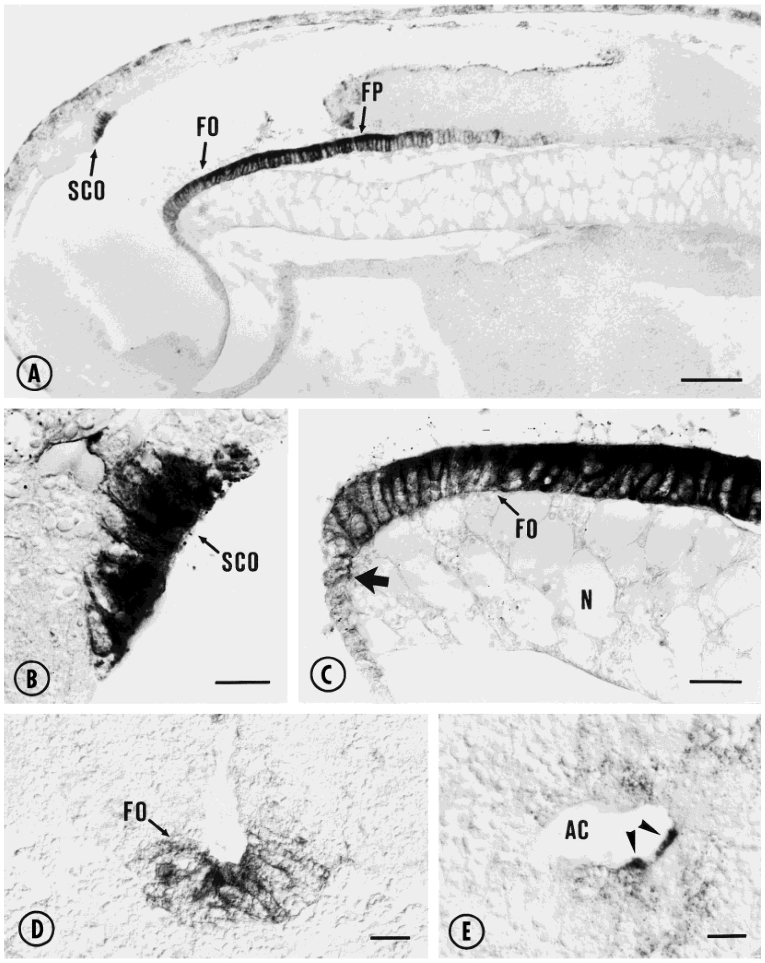
Fig. 6. Immunostaining of the CNS of Xenopus embryos using
AFRU. A: Midsagittal section of a 41-hour-old embryo. The immunocytochemical
reaction is confined to the SCO, FO, and FP. B: Higher
magnification of the SCO shown in a. C: Higher magnification of the
FO shown in a. AFRU-ir cells of the FO form a column closely apposed
to the notochord. The arrow indicates cephalic end of the immunoreactive
column of FP cells. D: Frontal section of a 33-hour-old embryo.
Moderate RF-immunoreactivity is seen in cells of the FO. E: Frontal
section of a 33-hour-old embryo. AFRU-ir material is present in the
lumen of the ampulla caudalis (arrowheads). Scale bar 5 30 μm. AC,
Ampulla caudalis; FO, flexural organ; FP, floor plate; N, notochord;
SCO, subcommissural organ. Scale bars5200 μm in A, 30 μm in B and
E, 50 μm in C and D. Image published in: Yulis CR et al. (1998) Copyright © 1998. Image reproduced with permission of the Publisher, John Wiley & Sons.
Image source: Published Larger Image Printer Friendly View |