XB-IMG-82721
Xenbase Image ID: 82721
|
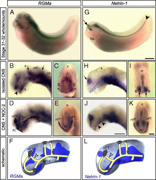
Fig. 5. The Neogenin ligands RGMa and Netrin-1 are expressed in the developing Xenopus forebrain. In situ hybridization for RGMa (A�E) and Netrin-1 (G�K) was performed on whole-mount stage 30�31 embryos (A, G) and isolated stage 30�31 brains, indicated by dashed lines (B�C, H�I). All staining is bilaterally symmetrical. Combined in situ staining (blue) with immunostaining for the NOC-2+ subset of forebrain axons (brown) was performed to show the relationship between axon tract formation and mRNA expression (D�E, J�K). (A) In lateral view of a whole-mount Xenopus embryo, RGMa expression is detected in the brain, spinal cord somites and branchial arches (ba). (B) In lateral view of an isolated brain, strong RGMa expression is detected in the forebrain (fb), midbrain (mb) and hindbrain (hb). Expression extends rostrally in a band from the dorsal telencephalon, terminating in a ventral �wedge� (asterisk) at the rostral end of the forebrain. RGMa expression is absent from the trajectory of the TPOC (arrowhead) but a patch of expression is detected in the ventral forebrain, dorsal to the hypothalamus (h). RGMa is expressed throughout the ventral half of the midbrain and hindbrain, extending in dorsal peaks at the forebrain�midbrain boundary and midbrain�hindbrain boundary (arrows). (C) In dorsorostral view (epiphysis (e) indicates the dorsal surface of the brain), RGMa expression in the forebrain is detected along the midline and in the neuroepithelium medial to the estimated position of the nPT (hatched circles, compare to double-labeled image in panel E). The rostroventral wedge of RGMa expression in the forebrain (asterisks) extends laterally. Lateral (D) and dorsorostral views (E) of brains double-labeled for NOC-2 shows that RGMa expression is absent from the pathway underlying the nPT, AC and TPOC. The SOT forms along the border of the rostroventral �wedge� of RGMa expression, then grows into a region in devoid of RGMa (unfilled arrowhead in panel D). (G) In lateral view of a whole-mount Xenopus embryo, Netrin-1 is detected in a distinct patch in the brain (arrow) and throughout the ventral CNS (arrowhead). Expression is also detected in the ventral retina and the branchial arches (ba). (H) Netrin-1 expression is strong throughout the ventral forebrain, midbrain and hindbrain. A distinct patch of expression is also present in the dorsal forebrain (arrow). (I) A dorsorostral view shows that Netrin-1 expression in the dorsal forebrain (arrows) is located medially, close to the midline. The estimated positions of the nPT are indicated (hatched circles, compare to double-labeled image in panel K). The epiphysis (e) denotes the dorsal surface of the brain. (I) Double-labeling in lateral (J) and dorsorostral view (K) shows that Netrin-1 expression lies medial to the nPT, and underlies the TPOC (arrowheads). (F, L) Schematic representations of the expression of RGMa and Netrin-1 relative to the axon scaffold is consistent with their potential interaction with Neogenin as ligand-receptor pairs in the guidance of nPT axons to form the SOT (unfilled arrowhead). Scale bar in panel G = 500 μm and also applies to panel A, scale bar in panel J = 60 μm and applies to panels B, D, H, scale bar in panel K = 60 μm and applies to panels C, E, I. Image published in: Wilson NH and Key B (2006) Copyright © 2006. Image reproduced with permission of the Publisher, Elsevier B. V.
Image source: Published Larger Image Printer Friendly View |