XB-IMG-80081
Xenbase Image ID: 80081
|
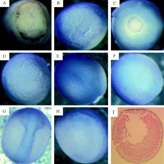
Fig. 1. Whole mount in situ analysis of the expression of Xlfc (blue/purple staining). (A,B) Stage 10 (early gastrula) embryos. (A) View of blastopore end, dorsal side lower most. (B) Lateral view, blastopore end to right. (C,D) Stage 12 (late gastrula) embryos. (C) View of blastopore end (posterior). Note even distribution of staining ventral and dorsal. (D) Anterior view. (E,F) Stage 14 (neurula) embryos. (E) Dorsal view, anterior top right; posterior bottom left. Note staining becoming concentrated in the dorsal (neural) ectoderm. (F) Ventral view. (G,H) Stage 18 (late neurula) stage. (G) Dorsal view, anterior top. Staining is now predominantly in the neural tube. (H) Lateral view, dorsal to right. (I) A 10-mm central transversal section through a late gastrula stage (12) albino embryo. The dorsal side is uppermost. Note that only the outer (ectodermal) cells are stained (brown). The section has been counter-stained red. Image published in: Morgan R et al. (1999) Copyright © 1999. Image reproduced with permission of the Publisher, Elsevier B. V.
Image source: Published Larger Image Printer Friendly View |