XB-IMG-44435
Xenbase Image ID: 44435
|
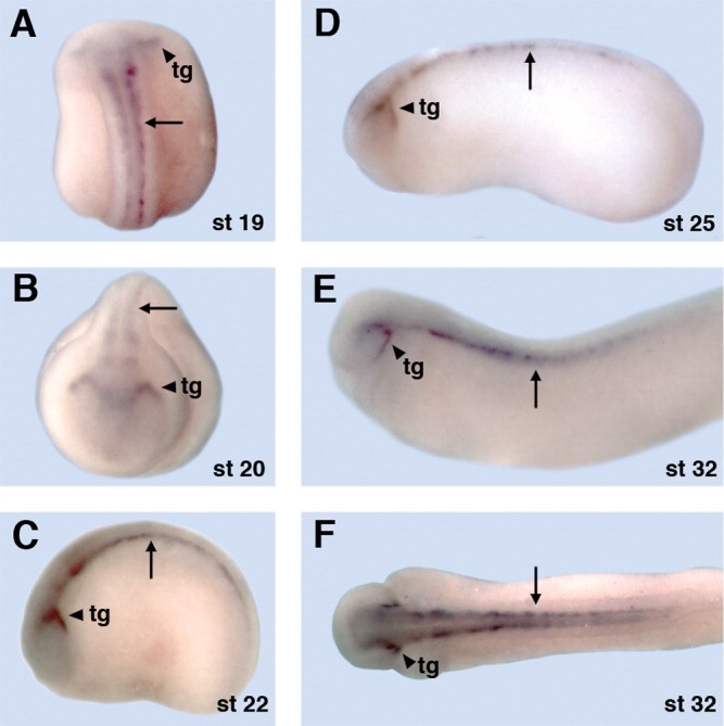
Figure 4. Developmental expression pattern of Xenopus SVOP. A-F: The expression pattern of SVOP was determined by performing whole-mount in situ hybridization on wild-type albino embryos. Arrowheads point to SVOP staining in the trigeminal ganglion (tg), whereas arrows indicate spinal cord expression. Embryo orientations are dorsal in A and F, anterior in B and lateral in C-E. SVOP, SVtwO-related Protein. Image published in: Logan MA et al. (2005) Copyright © 2005. Image reproduced with permission of the Publisher, John Wiley & Sons.
Image source: Published Larger Image Printer Friendly View |