XB-IMG-43995
Xenbase Image ID: 43995
|
||||||||||||||||||||||||||||||
|
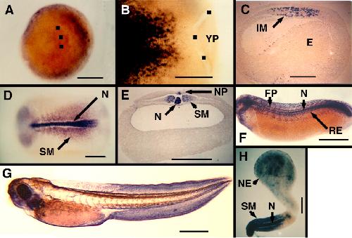
Figure 3. Localization of XF message in the embryo. XF message is detected by whole mount in situ hybridization. Dorsal is to the right in A, and is up in C, E, F, and G. Anterior is to the left in B, C, D, F, and G. A: Vegetal view of a St-10.5 embryo. Black squares are to the left of the blastopore lip. B: Dorsal view of the posterior half of St-11.5 whole mount. Black squares identify the blastopore lip. C: Sagittal section through a St-11.5 whole mount. D: Dorsal view of a St-16 embryo. E: Cross-section through the trunk of a St-16 embryo. F: Lateral view of St-25 embryo. G: Lateral view of St-40 embryo. H: Keller sandwich (St 20) with neural axis running anterior to posterior from top, and mesodermal axis running anterior to posterior from bottom, meeting at middle bend. YP, Yolk plug; N, notochord; SM- Somitic mesoderm; NP, notoplate; RE, roof of endoderm; NT, neural tube; IM, involuting mesoderm; E, endoderm; EB, eye bud; NE, neural extension. Scale bars = 0.25 mm (B-E), 0.5 mm (A,H), 1 mm (F,G). Image published in: Skoglund P et al. (2006) Copyright © 2006. Image reproduced with permission of the Publisher, John Wiley & Sons.
Image source: Published Larger Image Printer Friendly View |