XB-IMG-43557
Xenbase Image ID: 43557
|
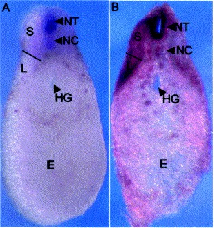
Fig. 2. A cross-section through the posterior end of a tail bud stage (26) embryo across the plane indicated in Fig. 1D. Dorsal is top and ventral is bottom. (A) XHoxb-4 staining. (B) XHoxc-8 staining. The line marks the division between the somite (S) and the lateral plate mesoderm (L). E, endodermal yolk mass; HG, hind gut; NT, neural tube; NC, notochord. Image published in: Thickett C and Morgan R (2002) Copyright © 2002. Image reproduced with permission of the Publisher, Elsevier B. V.
Image source: Published Larger Image Printer Friendly View |