Click here to close Hello! We notice that you are using Internet Explorer, which is not supported by Xenbase and may cause the site to display incorrectly. We suggest using a current version of Chrome, FireFox, or Safari.
XB-IMG-192685

Xenbase Image ID: 192685


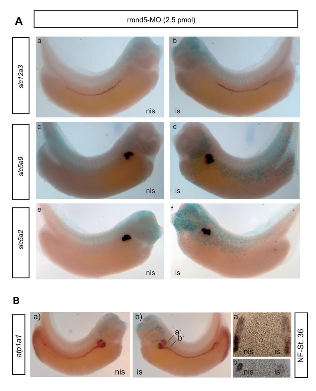
Fig. S3. Suppression of RMND5A function is not associated with defects in the segmentation of pronephric kidney during development. (A) rmnd5a- morpholino (2.5 pmol/embryo) injected embryos (NF st.36) were subjected to in situ hybridization with indicated marker probes; slc12a3 (a, b) and slc5a9 (c, d), and slc5a2 (e, f). (B) rmnd5a-morpholinos injected embryos were used for in situ hybridizations (WMISH) (NF36) with indicated marker atp1a1. a’ and b’ show zoom sections of embryos kidneys. Abbreviations: is, injected side; nis, non-injected side.

Image published in: Hantel F et al. (2022)

Copyright © 2022. Image reproduced with permission of the Publisher, The Company of Biologists Ltd.

GeneSynonymsSpeciesStage(s)Tissue
slc5a9.Lsglt1k, sglt4, XSGLT1KX. laevisThroughout NF stage 35 and 36pronephric kidney
pronephric nephrostome
proximal tubule
slc5a2.LX. laevisThroughout NF stage 35 and 36pronephric kidney
pronephric nephrostome
proximal tubule
slc12a3.Lncc, ncct, tscX. laevisThroughout NF stage 35 and 36pronephric duct
atp1a1.Latp1a1-a, atp1a1-b, K-ATPase, Na+/K+ ATPase, Na+K+ ATPase, Na+K+ATPaseX. laevisThroughout NF stage 35 and 36kidney
pronephric kidney
proximal tubule
pronephric nephrostome
pronephric duct
pronephric nephron
glomus
distal tubule

Image source: Published

Larger Image
Printer Friendly View

Return to previous page