XB-IMG-192525
Xenbase Image ID: 192525
|
||||||||||
|
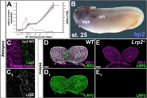
Fig. S1: Lrp2 expression and protein depletion by knock-down in Xenopus and knockout
in mouse. (A) mRNA and protein expression profile of Xenopus lrp2.L (from Xenbase.org).
(B) Xenopus lrp2 expressed in brain (arrowheads), eye anlage, otic vesicle (ov) and proximal
pronephros (pn) in stage (st.) 25 tailbud embryo. (C) Morpholino oligomer (MO) decreases
Lrp2 expression in injected cells; single channel (C1) for clarity. (D, E) Frontal views of anterior
neural folds of wild type (WT; D) and Lrp2-/- (E) mouse embryos at embryonic day (E) 8.5. (D1,
E1) WT LRP2 expression (D1) lost in Lrp2-/- (E1). ZO-1 labels cell boundaries. Scale bars (D,
E): 50 μm. Image published in: Kowalczyk I et al. (2021) Copyright © 2021. Image reproduced with permission of the publisher and the copyright holder. This is an Open Access article distributed under the terms of the Creative Commons Attribution License.
Image source: Published Larger Image Printer Friendly View |