XB-IMG-175731
Xenbase Image ID: 175731
|
||||||||||||||||||||||||||||||
|
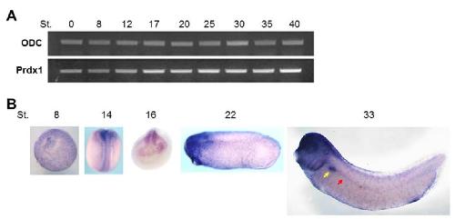
Figure S1. Spatiotemporal expression pattern of Prdx1 during embryogenesis.
A. Xenopus embryos were harvested at various stages and RT-PCR was performed using standard methods. The Numbers indicate the embryonic stages. odc was used as the loading control. The expression of prdx1, a maternal gene, gradually increased from the blastula to
the tadpole stage.
B. Whole mount in situ hybridization with a digoxigenin labeled antisense probe against prdx1
was performed for embryos at stage 8, 14, 16, 22 and 33. prdx1 was expressed in the forebrain, eye, multiciliated cells, and pronephros. The yellow arrow points to the developing pronephros; the red arrow points to the presumptive pronephric tubule ducts. Image published in: Chae S et al. (2017) © The Author(s) 2017. Creative Commons Attribution license
Image source: Published Larger Image Printer Friendly View |