XB-IMG-174472
Xenbase Image ID: 174472
|
|||||||||||||||||||||||||
|
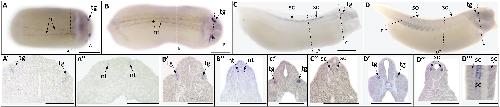
Fig. 1. Whole mount (A–D) and histological (A′-D‴) expression of trpv1 transcripts in developing X. laevis embryos. (A–B) dorsal view, (C–D) lateral view, anterior to the right for all whole mount embryos; dorsal to the top for all histology images. (A, A′, A′) stage 20 (late neurula stage); (B, B′, B′) stage 25 (early tailbud stage); (C, C′, C′) stage 30 (late tailbud stage); (D, D′, D′, D‴) stage 35 (swimming tadpole stage). Arrows indicate regions of gene expression (nt, nural tube; sc, spinal cord; tg, trigeminal ganglia). Dashed lines represent positions of corresponding sections. Scale bars = 250 μm. Image published in: Dong C et al. (2018) Copyright © 2018. Image reproduced with permission of the Publisher, Elsevier B. V.
Image source: Published Larger Image Printer Friendly View |