XB-IMG-171687
Xenbase Image ID: 171687
|
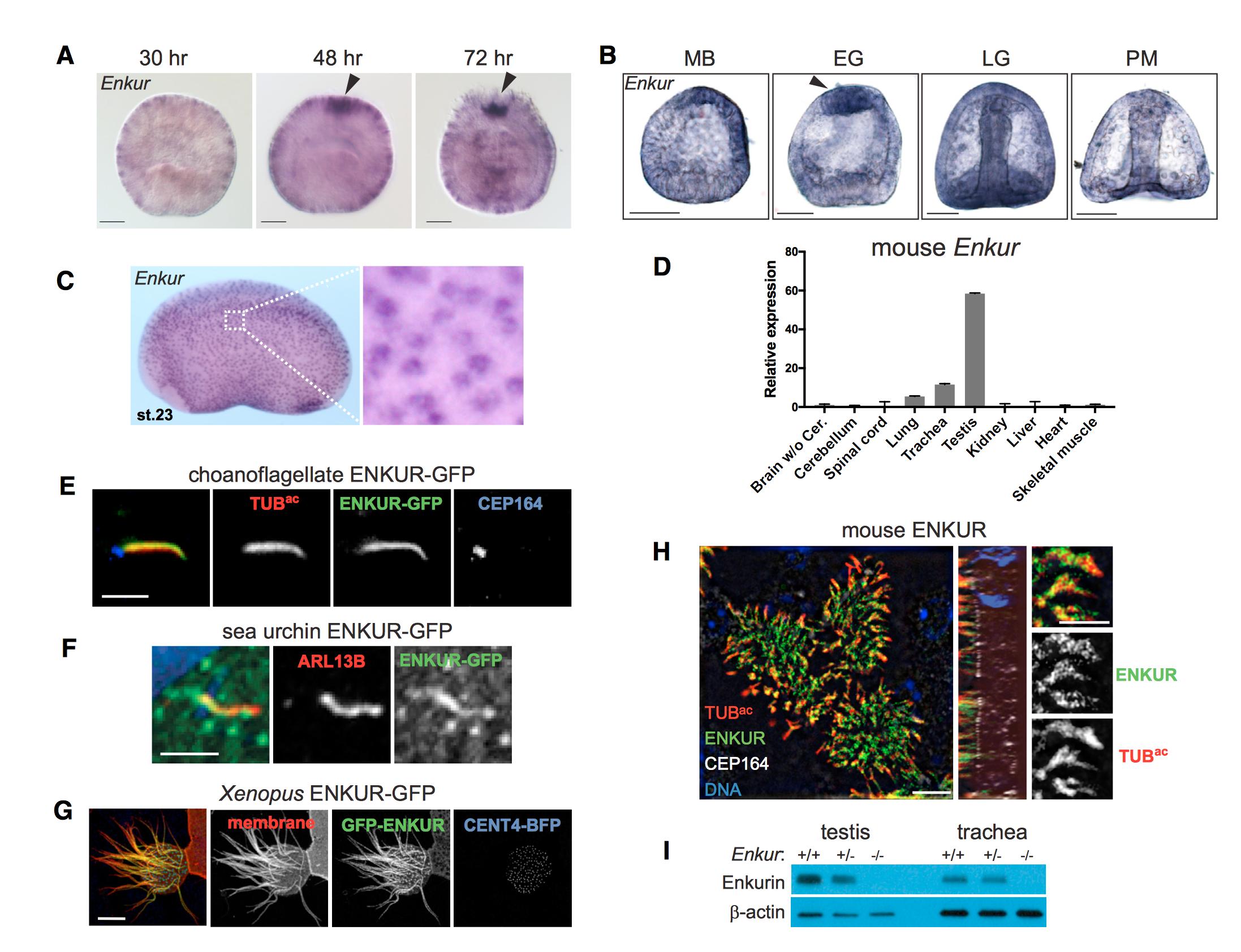
Figure 5. ENKUR Is a Conserved Ciliary Protein Expressed by Cells with Motile Cilia
(A) Whole-mount in situ hybridization for Enkur in N. vectensis embryos at various developmental stages. Enkur is expressed throughout the embryo and is enriched at the aboral pole (arrowhead) in 48- and 74-hr embryos. Scale bar, 50 mm.
(B) In situ hybridization for Enkur in S. purpuratus embryos at mesenchyme blastula (MB), early gastrula (EG), late gastrula (LG), and prism (PM) stages. Scale bar, 50 mm. Enkur is expressed in all cells and enriched at the apical pole (arrowhead) in EG embryos.
(C) In situ hybridization of stage-23 Xenopus laevis embryo shows expression of Enkur in epidermal cells.
(D) qRT-PCR measurement of Enkur detected expression in isolated adult mouse lungs, trachea, and testis. Error bars represent SDs from 6 technical replicates. Expression was validated using 2 distinct primer pairs.
(E) Immunofluorescent staining of S. rosetta ENKUR fused to GFP (green), cilia (TUBac, red), and the basal body (CEP164, blue) expressed in IMCD3 cells. Scale bar, 2.5 mm.
(F) Immunofluorescent staining of cilia (ARL13B, red) and a fusion of S. purpuratus ENKUR with GFP (green) expressed in RPE-1 cells. ENKUR-GFP localizes to cilia. Nuclei are stained with Hoechst (blue). Scale bar, 2.5 mm.
(G) A multiciliated epidermal cell of a stage-23 X. laevis embryo expressing X. laevis ENKUR fused to GFP (green), membrane-red fluorescent protein (red), marking the plasma and ciliary membranes, and Centrin 4 (CENT4)-blue fluorescent protein (BFP, blue) to mark the basal bodies. ENKUR localizes along the length of cilia. Scale bar, 10 mm.
(H) Immunofluorescent staining of primary cultured mouse tracheal epithelial cells for ENKUR (green), cilia (TUBac, red), and basal bodies (CEP164, white), and imaged using structured illumination microscopy. ENKUR localizes to mouse tracheal epithelial cilia. Scale bar for whole cells (top and center panels), 5 mm. Scale bar for cilia (right panel), 2.5 mm.
(I) Immunoblotting for ENKUR protein in testis and trachea lysates from littermate control and Enkur -/- mice. ENKUR is expressed in testis and trachea, whereas Enkur -/- mice do not produce detectable ENKUR.
See also Figures S4 and S5. Image published in: Sigg MA et al. (2017) Copyright © 2017. Image reproduced with permission of the Publisher, Elsevier B. V.
Image source: Published Larger Image Printer Friendly View |