XB-IMG-171463
Xenbase Image ID: 171463
|
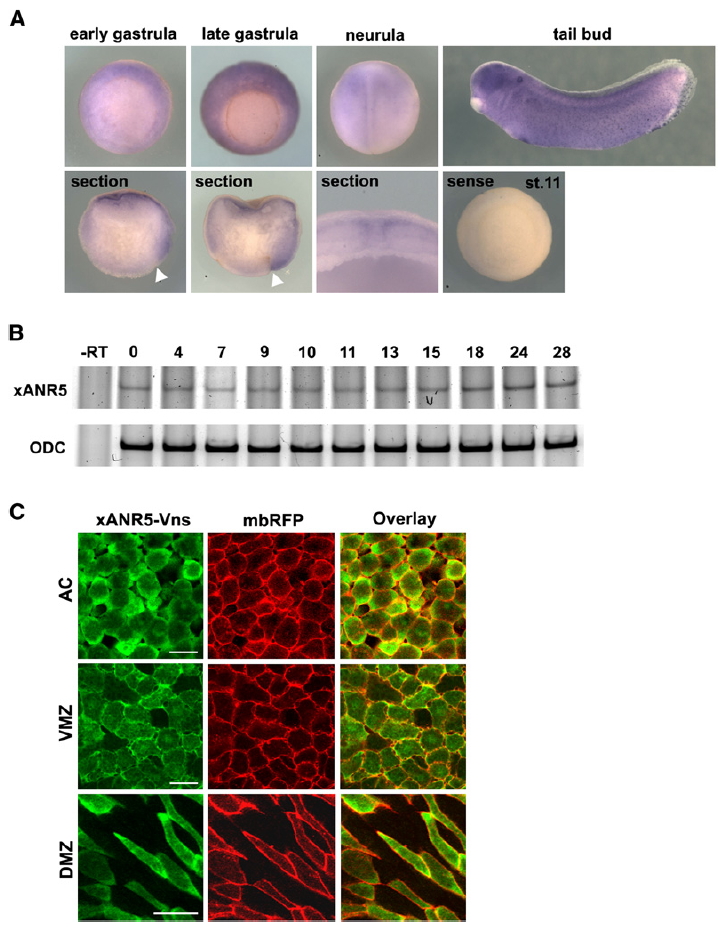
Figure S1. Expression Pattern and Subcellular Localizations of xANR5 through Early Embryogenesis (A and B) Whole-mount in situ hybridization analysis (WISH) and semiquantitative RT-PCR analyses showed ANR5 to be the product of a maternally expressed gene. The expression patterns of xANR5 in the gastrula were almost ubiquitous but dorsally enriched, as seen in the sagittal section. xANR5 RNA was expressed along the neural fold in the neurula, and observed in the eyes, midbrain, and hindbrain, and as dots in the epidermis in later stages. (C) Subcellular localization of xANR5 during early embryogenesis was detected by means of a Vns-tagged construct. A membrane-bound form of RFP was coinjected to detect plasma membrane. xANR5-Vns was localized near the plasma membrane in the AC and VMZ. In the DMZ, xANR5-Vns was localized to the two tips of the spindle-shaped dorsal cells. Scale bars represent 50 mm. Image published in: Chung HA et al. (2007) Copyright © 2007. Image reproduced with permission of the Publisher, Elsevier B. V.
Image source: Published Larger Image Printer Friendly View |