XB-IMG-144792
Xenbase Image ID: 144792
|
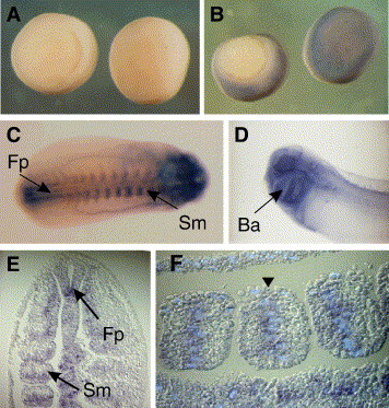
Fig. 1. SMRT is expressed predominantly in the head and somites of the tailbud Xenopus embryo. (A, B) In situ hybridization of gastrula stage embryos using (A) sense and (B) antisense SMRT RNA probes. (C) Dorsal view of a wholemount in situ hybridization of an early tailbud embryo (stage 26) using an antisense SMRT probe (anterior to right). There is significant staining in the head and posterior floorplate (Fp). Lateral stripes correspond to the position of the somites (Sm) and expression is also seen in the posterior mesoderm that has yet to segment. (D) In the late tailbud (stage 32) embryo, expression is predominantly within the head in the branchial arches (Ba) and eyes. (E) Planar longitudinal section through a tailbud embryo (posterior to the top) showing the expression of SMRT in the somites (Sm) and in the floor plate (Fp) of the posterior neural tube. (F) Highlighting the nuclei within the cells of a somite using Hoechst stain shows that SMRT mRNA has a perinuclear distribution (arrowed). Image published in: Malartre M et al. (2006) Copyright © 2006. Image reproduced with permission of the Publisher, Elsevier B. V.
Image source: Published Larger Image Printer Friendly View |