XB-IMG-138279
Xenbase Image ID: 138279
|
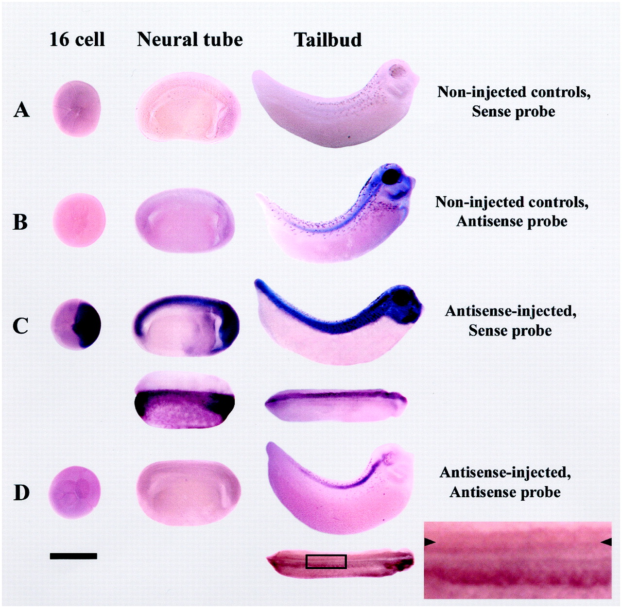
Fig. 2.
Inhibition of expression of xKv3.1transcripts by antisense RNA injection is visualized by in situ hybridization of whole-mount Xenopus embryos with a xKv3.1-specific antisense probe. A, Sense (S) probe gives no signal in uninjected control embryos. B, AS probe reveals that endogenous xKv3.1 mRNA is present in the spinal cord of control embryos at stage 22 and thereafter (Gurantz et al., 2000).C, S probe labeling of AS-injected embryos demonstrates stability of injected xKv3.1 AS. Signal is observed in 16-cell through stage 30 embryos; dorsal view of another embryo indicates that only the injected side of the embryo (top) contains AS. D, Injection of xKv3.1 AS reduces the endogenous signal detected with the AS probe. Dorsal view of another embryo indicates that reduction of the in situ hybridization signal is restricted to the injected side (top, arrowheads). Area inbox is enlarged at right. For all embryos anterior is to the right and dorsal isup. Scale bar, 1 mm. Black spots on tailbud embryos are melanocytes. Image published in: Vincent A et al. (2000) Copyright © 2000. This image is reproduced with permission of the publisher and the copyright holder. This is an Open Access article distributed under the terms of the Creative Commons Attribution License.
Image source: Published Larger Image Printer Friendly View |