XB-IMG-135141
Xenbase Image ID: 135141
|
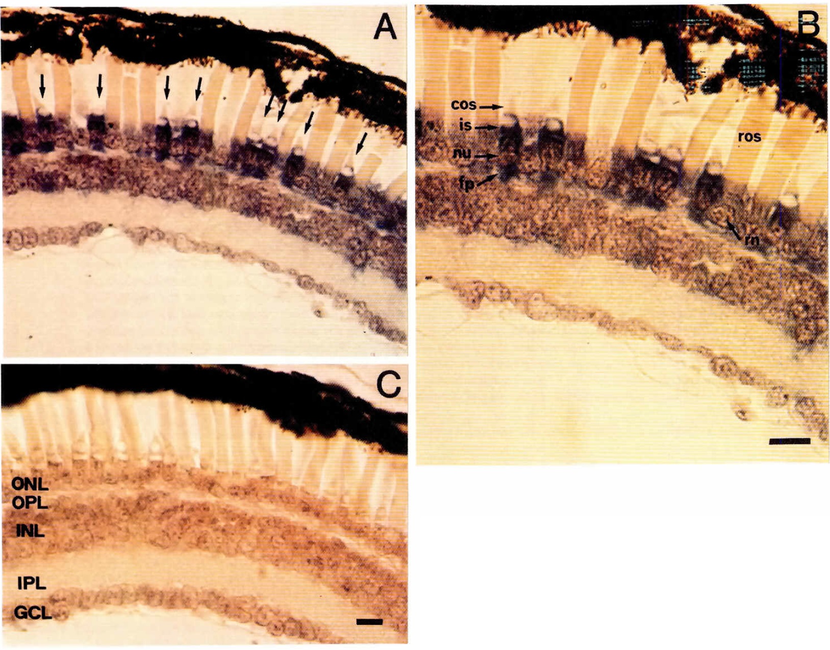
Fig. 1. Immunostaining analysis of retinal Xenopus swimming tadpole
sections. stained with the anti-Xfin antibody. (AI The arrows point
to the stained cones. (BI Enlargement of panelA. Cos: cone outer segment;
ros: rod outer segment; nu: nucleus, fp: footpiece. IC) Negative control made
by using preimmune serum. ONL: outer nuclear layer; OPL: outer plexiform
layer; INL: inner nuclear layer; IPL: inner plexiform layer; GCL: ganglion cel!
layer. Bars represent 5um. Image published in: Rijli FM et al. (1993) Copyright © 1993. Reproduced with permission of the Publisher, University of the Basque Country Press.
Image source: Published Larger Image Printer Friendly View |