Click here to close Hello! We notice that you are using Internet Explorer, which is not supported by Xenbase and may cause the site to display incorrectly. We suggest using a current version of Chrome, FireFox, or Safari.
XB-IMG-133902

Xenbase Image ID: 133902


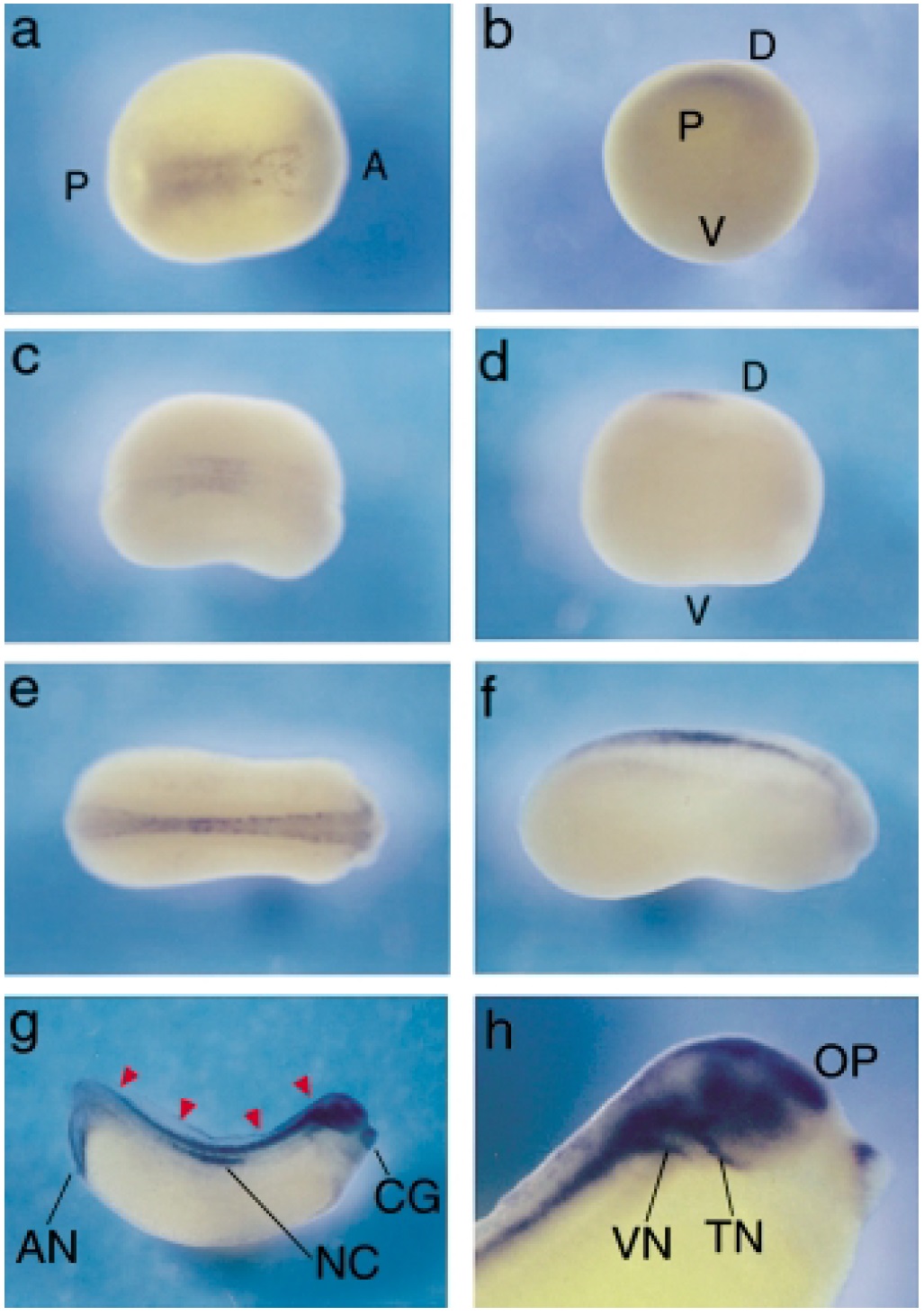
Fig. 5. Whole-mount in situ hybridization. Expression pattern of Xp8 at stages (a, b) 14, (c, d) 18, (e, f) 22 and (g, h) 30. (a,c,e) Dorsal (D) view, (d,f–h) lateral view, (b) ventroposterior view. The anterior (A) is situated on the right side in all parts except (b). Arrowheads show the central nervous system. V, ventral; P, posterior; AN, anus; NC, notochord; CG, cement gland; VN, vestibulocochlear nerve; TN, trigeminal nerve; OP, olfactory placode.

Image published in: Igarashi T et al. (2001)

Copyright © 2001. Image reproduced with permission of the Publisher, John Wiley & Sons.

GeneSynonymsSpeciesStage(s)Tissue
nupr1.Lcom1, p8, Xp8X. laevisThroughout NF stage 14 to NF stage 18neural plate
chordal neural plate
nupr1.Lcom1, p8, Xp8X. laevisThroughout NF stage 22neural tube
posterior neural tube
anterior neural tube
nupr1.Lcom1, p8, Xp8X. laevisThroughout NF stage 29 and 30proctodeum
trigeminal nerve
cement gland
cranial nerve
vestibulocochlear ganglion
brain
forebrain
olfactory placode
notochord
spinal cord
cloaca
central nervous system
nerve

Image source: Published

Larger Image
Printer Friendly View

Return to previous page