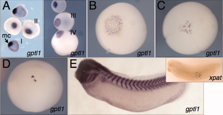
Figure 4. Expression and germ plasm localization of gptl1. In situ hybridization against gptl1. A: Early stage oocytes (stage I-II, left panel; stage III-IV, right panel). Roman numerals indicate oocyte stages; mc, mitochondrial cloud. B: Stage VI oocyte, vegetal view. C: Four-cell embryo, vegetal view. D: Stage 10.5, vegetal view. E: Stage 30 embryo, cleared in Murray's clear, showing expression in branchial arches and intersomitic boundaries and the absence of staining in primordial germ cells (PGCs). Inset: in situ for xpat showing positive PGC staining in a cleared embryo.
Image published in: Cuykendall TN and Houston DW (2010)
Copyright © 2010. Image reproduced with permission of the Publisher, John Wiley & Sons.
| Gene | Synonyms | Species | Stage(s) | Tissue |
|---|---|---|---|---|
| pes1.S | germ plasm transcribed locus 1, gptl1, pes, pescadillo | X. laevis | Throughout NF stage 10.5 | vegetal pole germ plasm |
| pes1.S | germ plasm transcribed locus 1, gptl1, pes, pescadillo | X. laevis | Throughout NF stage 29 and 30 | eye pharyngeal arch central nervous system spinal cord brain forebrain midbrain hindbrain mandibular arch hyoid arch branchial arch pronephric tubule otic vesicle |
| pgat.L | Xpat | X. laevis | Throughout NF stage 29 and 30 | primordial germ cell germ plasm |
| pes1.S | germ plasm transcribed locus 1, gptl1, pes, pescadillo | X. laevis | Throughout NF stage 3 (4-cell) | germ plasm vegetal vegetal pole |
| pes1.S | germ plasm transcribed locus 1, gptl1, pes, pescadillo | X. laevis | Throughout unfertilized egg stage | oocyte germ plasm stage VI oocyte vegetal vegetal pole zygote cortex stage III oocyte stage I oocyte mitochondrial cloud stage IV oocyte |
Image source: Published
Permanent Image Page
Printer Friendly View
XB-IMG-42921