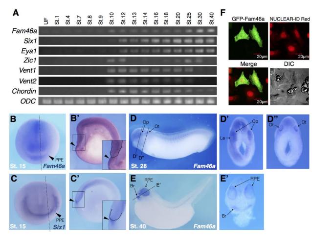
Fig. 1. Temporal and spatial expression pattern of Fam46a [tent5a] in Xenopus. (A) RT-PCR analysis of Fam46a expression. The expression of Fam46a is faintly detected from the maternal stage to stage 9, and increased from stage 10, similar to that of Zic1 (NPB), Vent1, Vent2 (BMP target gene), and Chordin (BMP antagonist). The expression of PPE marker genes, Six1 and Eya1, increased soon after the onset of Fam46a expression. ODC was used as a control. (B-E) Whole-mount in situ hybridization (WISH) was performed with a Fam46a (B, Bâ) and Six1 (C, Câ) probe. The anterior view (B, C); the lateral view (D, E); the hemi-section (Bâ, Câ, Dâ, Dâ, Eâ). Development ⢠Accepted manuscript The stage of the embryo was shown in each panel. PPE, pre-placodal ectoderm; Op, optic vesicle; Ot, otic vesicle; Le, lens; Br, branchial arch mesenchyme; RPE, retinal pigment epithelium. (F) HeLa cells were transfected with GFP-Fam46a and cultured for 24 hours. Nuclei were stained with NUCLEAR-ID Red. Scale bars represent 20 μ m.
Image published in: Watanabe T et al. (2018)
Copyright © 2018. Image reproduced with permission of the Publisher and the copyright holder. This is an Open Access article distributed under the terms of the Creative Commons Attribution License.
| Gene | Synonyms | Species | Stage(s) | Tissue |
|---|---|---|---|---|
| tent5a.L | fam46a | X. laevis | Throughout NF stage 15 | anterior placodal area eye primordium neural plate chordal neural plate |
| six1.L | XSix1 | X. laevis | Throughout NF stage 15 | preplacodal ectoderm neural plate |
| tent5a.L | fam46a | X. laevis | Throughout NF stage 28 | otic vesicle optic vesicle spinal cord |
| tent5a.L | fam46a | X. laevis | Throughout NF stage 40 | retinal pigmented epithelium branchial arch skeleton |
Image source: Published
Permanent Image Page
Printer Friendly View
XB-IMG-173784