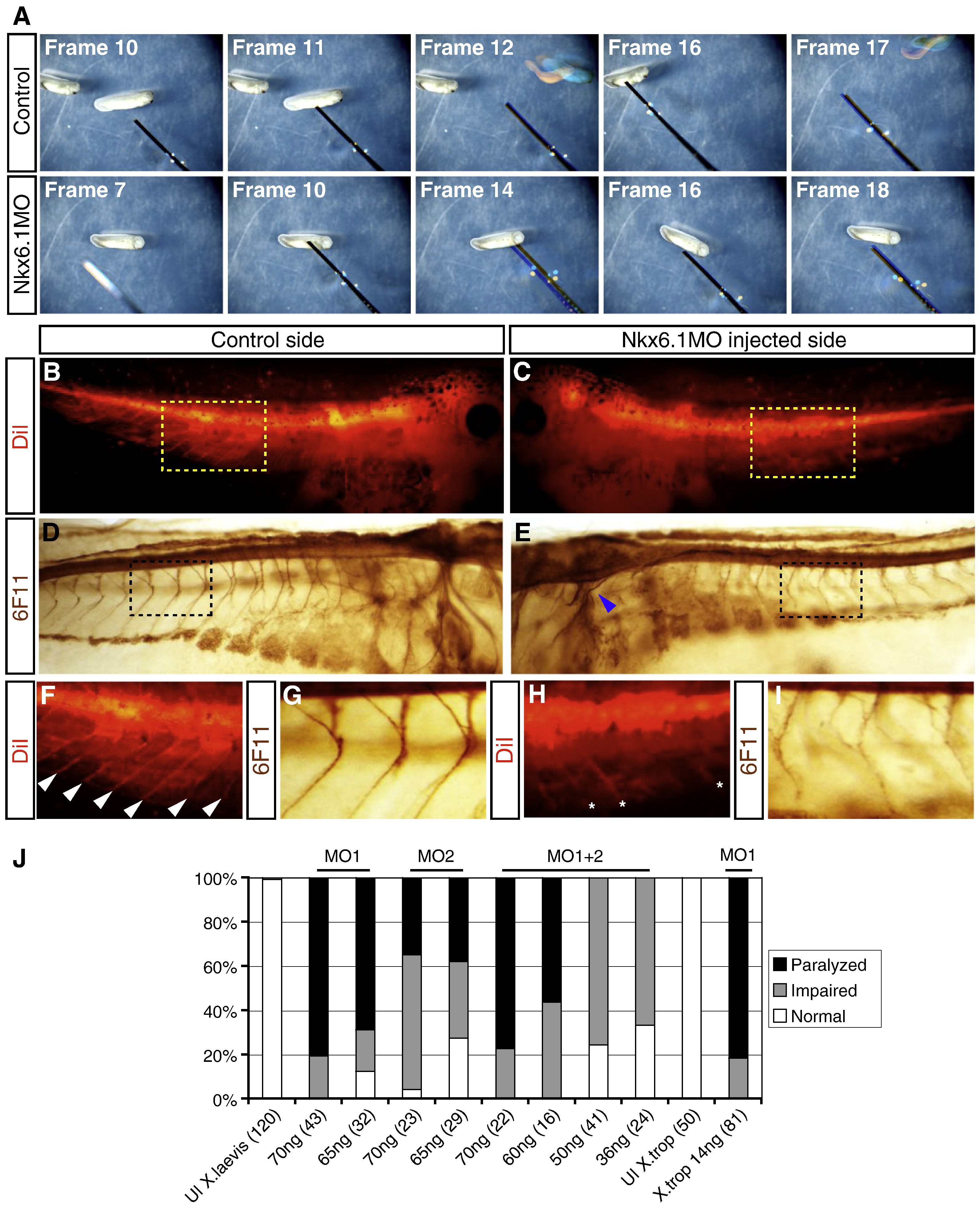
Fig.6. Nkx6.1 knockdown results in paralysis and axon misguidance. (A) Narrative frames from a time lapse series showing uninjected controls (top panel) and Nkx6.1MO injected embryos (bottom). Uninjected embryos initiate escape reflex when poked with a pipette tip (frames 12 and 17; pipette tip painted black). Poking of Nkx6.1MO injected embryos does not trigger escape reflex (bottom). Nkx6.1MO injected embryos were injected in both blastomeres at the 2-cell stage. (B,C,F,H) Orthograde DiI-labeling of stage 39/40 embryos injected unilaterally with Nkx6.1MO. (B) Control side showing labeled nerve fibers emanating from the neural tube. (C) Nkx6.1MO injected side show labeled nerves. (D,E,G,I) Immunostaining for N-CAM using antibody 6F11 showing axonal extensions. (D) Uninjected side of stage 39/40 tadpole showing nerve fibers protruding from the spinal cord and hindbrain. (E) Nkx6.1MO injected side of same tadpole does have greatly reduced and disorganized axons and cranial nerves are severely affected (blue arrowhead). (F, H) High magnification of boxes in (B) and (D), respectively. White arrow heads in (F) indicate DiI labeled axons. (H,I) High magnification of boxes show in (C) and (E), respectively. Asterisks in (H) indicate fluorescence from the uninjected side out of the plane of focus. (J) Effect of different MO1 and MO2 doses, individually and in combination, on tadpole mobility. Embryos were injected in all blastomeres at the one or two cell stage. Total MO amount injected is listed under bar. Number in parenthesis indicates number of embryos analyzed. MO1Â +Â 2 injections were performed with equal amounts of each. The two last bars show effect on MO1 when injected into X. tropicalis. Embryos were scored at stages 32â39.
Image published in: Dichmann DS and Harland RM (2011)
Copyright © 2011. Image reproduced with permission of the Publisher, Elsevier B. V.
Permanent Image Page
Printer Friendly View
XB-IMG-73853