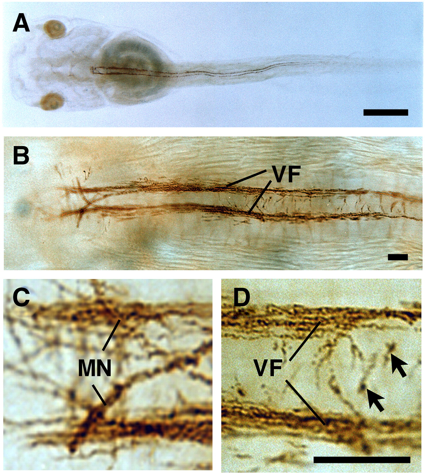
Fig. 6. Whole-mount immunocytochemistry of XMBP distribution in larvae. (AâD- Stage-47 albino larvae were immunocytochemically stained with the 3H8 Ab. MN, Mauthner neuron; VF, ventral fascicles of spinal cord. Arrows in D indicate immature myelin-forming cells. Scale bars in A, 1 mm; scale bars in B and D, 200 µm.
Image published in: Nanba R et al. (2010)
Copyright © 2010. Image reproduced with permission of the Publisher, Elsevier B. V.
| Gene | Synonyms | Species | Stage(s) | Tissue |
|---|---|---|---|---|
| mbp.L | xmbp | X. laevis | Throughout NF stage 47 | Mauthner cell spinal cord spinal neuron myelin accumulating cell |
Image source: Published
Permanent Image Page
Printer Friendly View
XB-IMG-180424