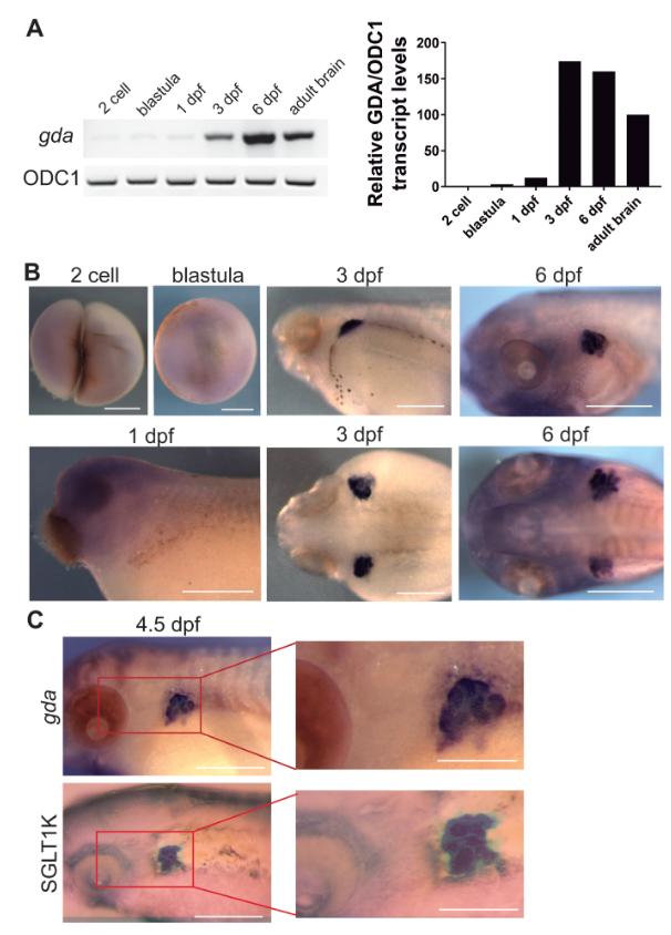
Figure 3. Xenopus GDA is expressed in the developing kidney.A. RT-PCR showing gdaexpression in the different developmental stages: 2 cell, blastula, neurula, 3 dpf, 6 dpf, and adult. ODC1 was used as an internal RT-PCR reference. B. Whole-mount in situ hybridization using an antisense probe specific to gda in different developmental stages. Scale bar: 0.5 mmC. Whole-mount in situ hybridization using an antisense probe specific to GDA or the kidney marker xSGLT-1K in 4.5 dpf embryos. Both gda and xSGLT-1K can be seen in a similar embryonic location. Scale bar: 0.5 mm and 0.25 mm for magnifications.
Image published in: Slater PG et al. (2019)
Copyright © 2019. Image reproduced with permission of the Publisher, John Wiley & Sons.
| Gene | Synonyms | Species | Stage(s) | Tissue |
|---|---|---|---|---|
| gda.L | X. laevis | Sometime during NF stage 41 to NF stage 45 | pronephric kidney | |
| gda.L | X. laevis | Sometime during NF stage 45 to NF stage 47 | pronephric kidney | |
| gda.L | X. laevis | Sometime during NF stage 47 to NF stage 48 | pronephric kidney | |
| slc5a9.L | sglt1k, sglt4, XSGLT1K | X. laevis | Sometime during NF stage 45 to NF stage 47 | pronephric kidney |
Image source: Published
Permanent Image Page
Printer Friendly View
XB-IMG-174837