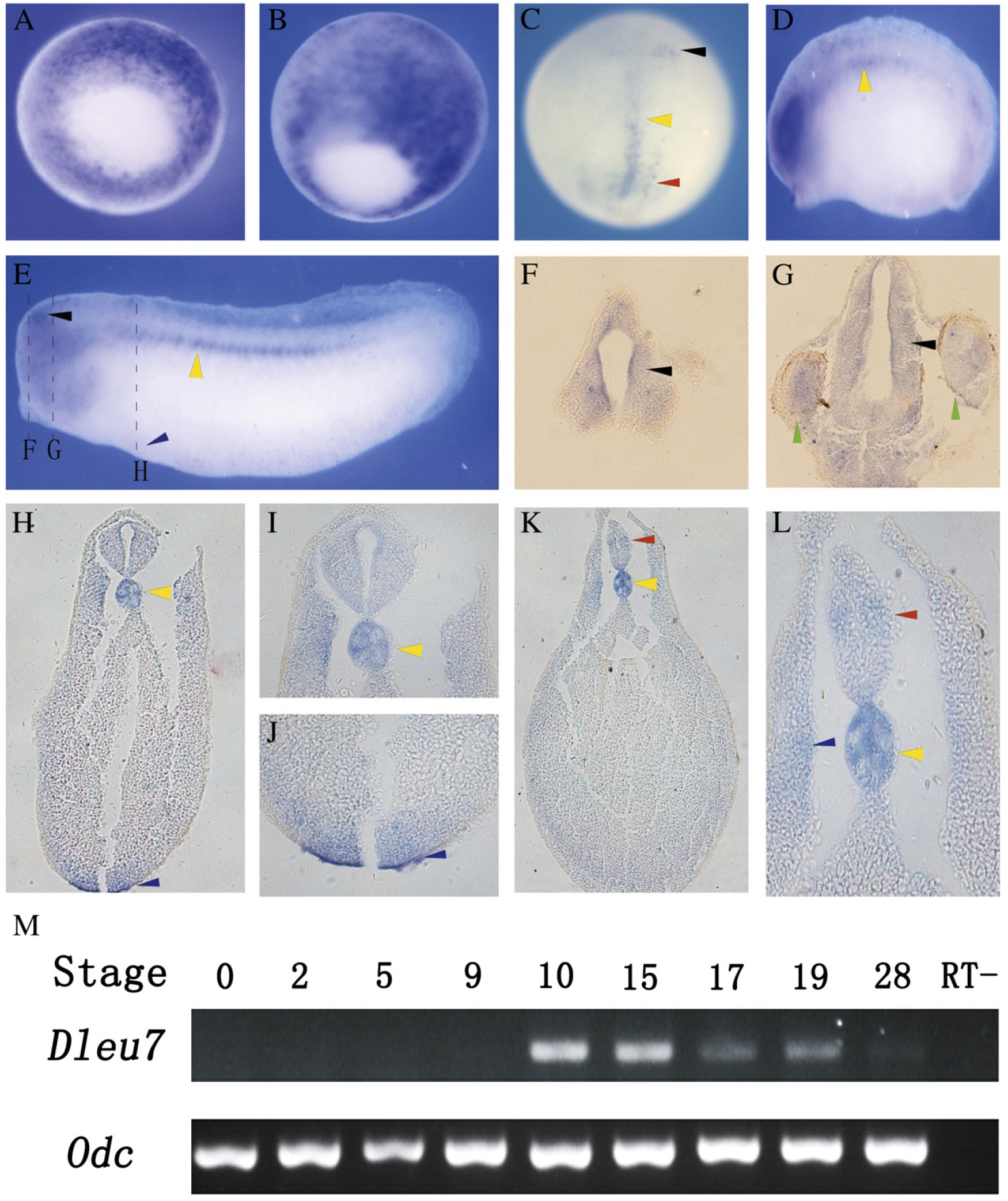
Fig. 6. Temporal and spatial expression of X. tropicalis Dleu7. (AâE) Spatial expression of Dleu7 by RT-PCR at stage 10.5 (gastrula) (A and B), stage 13 (early neurula) (C), stage 23 (late neurula) (D), and stage 28 (tail bud) (E). (FâL) Transverse paraffin section on stage 28 embryos after WISH. The signal of Dleu7 is observed in forehead (F), midbrain and eyes (G), notochord (I), blood island (H and J), neural tube and lateral muscle (K and L). The black, yellow, green, and red arrowheads indicate the signal of Dleu7 in brain, notochord, eye, and spinal cord respectively; the blue arrowheads show the expression of Dleu7 in the blood island (E, H) and lateral muscle (L). (M) Temporal expression of Dleu7. The expression is not detected from eggs to embryos until stage 9 and increases sharply at gastrula stage. The expression decreased gradually during the neurulation, and very weak signal is detected at stage 28 by RT-PCR.
Image published in: Zhu X et al. (2012)
Copyright © 2012. Image reproduced with permission of the Publisher, Elsevier B. V.
| Gene | Synonyms | Species | Stage(s) | Tissue |
|---|---|---|---|---|
| dleu7.L | MGC89906 | X. laevis | Sometime during NF stage 10.5 to NF stage 13 | mesoderm notochord marginal zone |
| dleu7.L | MGC89906 | X. laevis | Throughout NF stage 23 | notochord optic vesicle |
| dleu7.L | MGC89906 | X. laevis | Throughout NF stage 28 | notochord ventral blood island brain eye |
Image source: Published
Permanent Image Page
Printer Friendly View
XB-IMG-153751