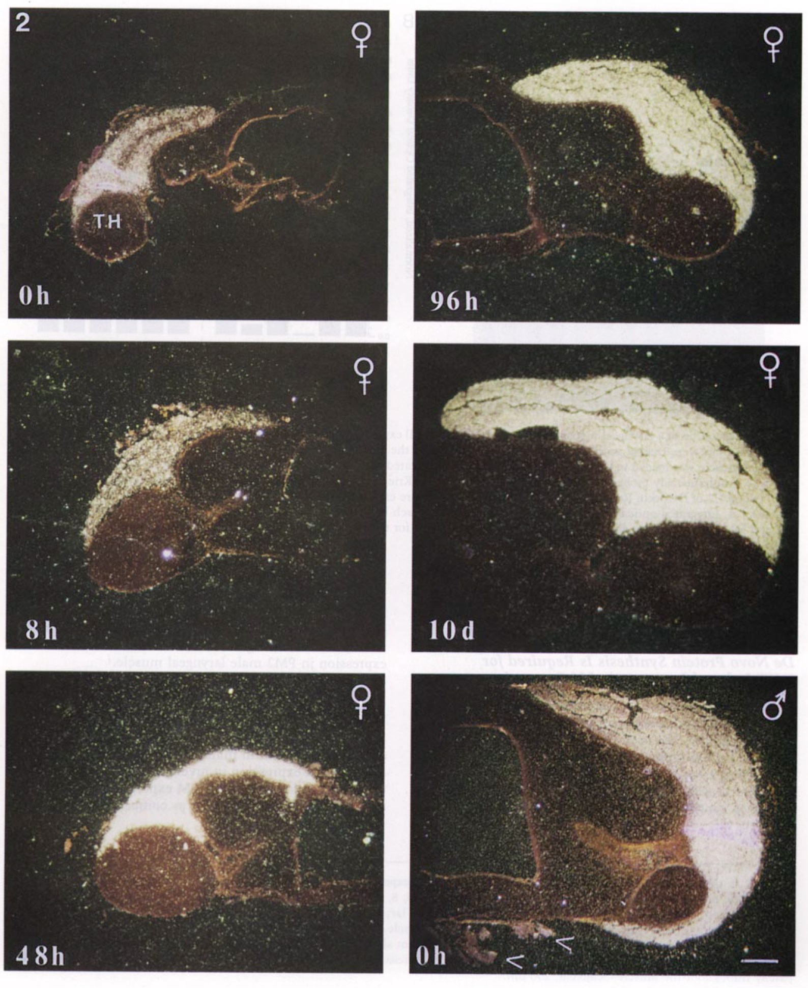
FIG. 2. In situ hybridization analysis of androgen-regulated LM expression. Dark-field micrographs of transverse 10·um hemisections through the larynges of female PM2 frogs treated with DHT for 0, 8, 48, 96, or 240 hr (240 hr = 10 days) are shown. For comparison,hybridization of an antisense LM probe to an untreated PM2 male larynx (panel at lower right, 0 hr) is also shown. The hybridization of the LM probe to an extrinsic muscle of the larynx, the strap muscle, did not exceed background !see arrowheads, male 0 hr), control hybridization with sense probes also did not exceed background (not shown). In the section of female larynx at 0 hr, a fold immediately above the thyohyral cartilages (TH) gives a false impression of increased expression relative to the rest of the muscle. LM expression is patchy throughout the muscle. Scale bar, 250 um.
Image published in: Catz DS et al. (1995)
Copyright © 1995. Image reproduced with permission of the Publisher, Elsevier B. V.
| Gene | Synonyms | Species | Stage(s) | Tissue |
|---|---|---|---|---|
| myh3.S | LM, LOC108701196 | X. laevis | Throughout juvenile frog stage | larynx laryngeal muscle |
Image source: Published
Permanent Image Page
Printer Friendly View
XB-IMG-145509