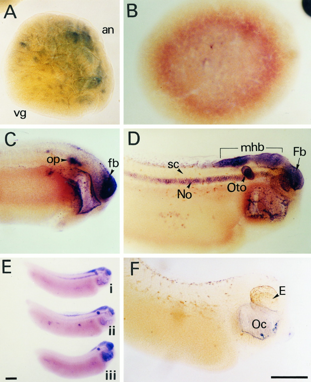
FIG. 5. Spatial distribution of Xrel3 mRNA during development. Albino embryos were subjected to whole mount in situ hybridization analysis using Xrel3- specific probes. The dark blue staining indicates localization of message. A, blastula; B, gastrula; C, late neurula; D and E, larva; F, control; op, otic placode; fb, forebrain; mhb, mid-hindbrain; oto, otocyst; No, notochord; sc, spinal cord; E, eye; Oc, oral cavity. Scale bar 5 0.5 mm.
Image published in: Yang S et al. (1998)
Copyright © 1998. Image reproduced with permission of the publisher and the copyright holder. This is an Open Access article distributed under the terms of the Creative Commons Attribution License.
| Gene | Synonyms | Species | Stage(s) | Tissue |
|---|---|---|---|---|
| rel.L | c-rel, rel2, rel3, v-rel, xrel, Xrel2, Xrel3 | X. laevis | Throughout NF stage 10 | animal hemisphere |
| rel.L | c-rel, rel2, rel3, v-rel, xrel, Xrel2, Xrel3 | X. laevis | Throughout NF stage 11 | |
| rel.L | c-rel, rel2, rel3, v-rel, xrel, Xrel2, Xrel3 | X. laevis | Throughout NF stage 15 to NF stage 22 | brain forebrain otic placode archenteron archenteron floor gastrocoel roof plate foregut dorsal endoderm anterior neural tube foregut endoderm liver diverticulum |
| rel.L | c-rel, rel2, rel3, v-rel, xrel, Xrel2, Xrel3 | X. laevis | Throughout NF stage 28 | notochord central nervous system spinal cord brain forebrain hindbrain midbrain-hindbrain boundary midbrain pharynx pharyngeal region pharyngeal arch otic vesicle |
Image source: Published
Permanent Image Page
Printer Friendly View
XB-IMG-135858