| Images | Sources | Experiment + Assay | Phenotypes | Human Diseases | ||
|---|---|---|---|---|---|---|

|
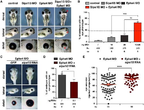
fig.5.a, b
Rothe M et al. (2017) |
Xla Wt + epha4 MO + sipa1l3 MO
NF42 (whole-mount microscopy) |
|
|||

|
fig.5.c, d, e
Rothe M et al. (2017) |
Xla Wt + Rno.sipa1l3 + epha4 MO
NF42 (whole-mount microscopy) |
|
|||

|
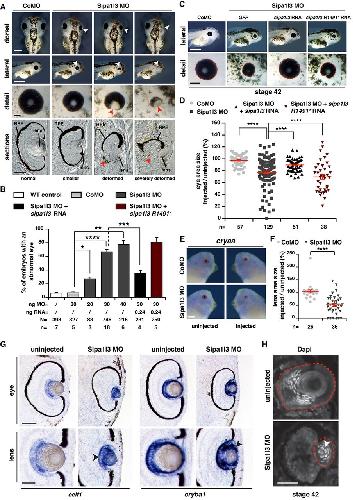
fig.2.b
Rothe M et al. (2017) |
Xla Wt + Rno.sipa1l3R1491* + sipa1l3 MO
NF42 (whole-mount microscopy) |
|
|||

|
fig.2.c, d
Rothe M et al. (2017) |
Xla Wt + Rno.sipa1l3R1491* + sipa1l3 MO
NF42 (whole-mount microscopy) |
|
|||

|
fig.2.a
Rothe M et al. (2017) |
Xla Wt + sipa1l3 MO
NF42 (whole-mount microscopy) |
||||

|
fig.2.b
Rothe M et al. (2017) |
Xla Wt + sipa1l3 MO
NF42 (whole-mount microscopy) |
|
|||

|
fig.2.c, d
Rothe M et al. (2017) |
Xla Wt + sipa1l3 MO
NF42 (whole-mount microscopy) |
|
|||

|
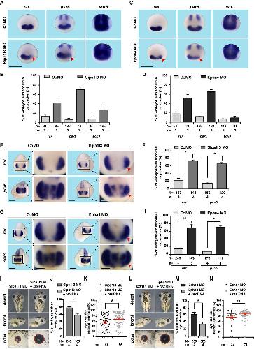
fig.6.i, j, k
Rothe M et al. (2017) |
Xla Wt + sipa1l3 MO
NF42 (whole-mount microscopy) |
|
|||

|
fig.6.l, m, n
Rothe M et al. (2017) |
Xla Wt + sipa1l3 MO
NF42 (whole-mount microscopy) |
|
|||

|
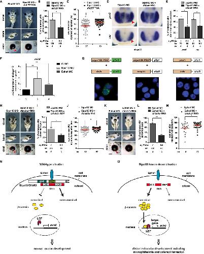
fig.7.a, b, c
Rothe M et al. (2017) |
Xla Wt + sipa1l3 MO
NF42 (whole-mount microscopy) |
|