abnormal development of eye has 7 anatomical phenotype(s)
Images Sources Experiment + Assay Phenotypes Human Diseases
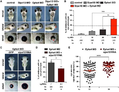
fig.5.a, b

Rothe M et al. (2017)
Xla Wt + epha4 MO + sipa1l3 MO

NF42 (whole-mount microscopy)
Anatomical Phenotype
abnormal development of eye
abnormal eye morphology
decreased size of the eye
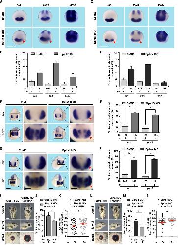
fig.6.i, j, k

Rothe M et al. (2017)
Xla Wt + sipa1l3 MO

NF42 (immunohistochemistry)
Anatomical Phenotype
abnormal development of eye
fig.6.l, m, n

Rothe M et al. (2017)
Xla Wt + sipa1l3 MO

NF42 (immunohistochemistry)
Anatomical Phenotype
abnormal development of eye
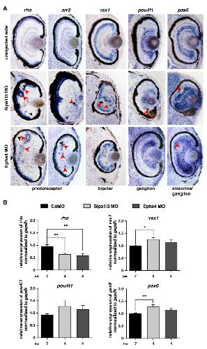
fig.S2.a

Rothe M et al. (2017)
Xla Wt + sipa1l3 MO

NF42 (in situ hybridization)
Anatomical Phenotype
abnormal development of eye
abnormal development of retinal layer
abnormal retinal ganglion cell layer morphology
abnormally localised retinal ganglion cell layer
abnormally localised retinal inner nuclear layer
Expression Phenotype
decreased amount rho.L expression in photoreceptor layer
increased amount pax6.L expression in amacrine cell
increased amount pax6.L expression in retinal ganglion cell layer
increased amount pou4f1.L expression in retinal ganglion cell layer
increased amount vsx1.L expression in retinal inner nuclear layer
mislocalised pax6.L expression in photoreceptor layer
mislocalised pax6.L expression in retinal inner nuclear layer
mislocalised rho.L expression in retinal inner nuclear layer
mislocalised vsx1.L expression in retinal ganglion cell layer
fig.5.a, b

Rothe M et al. (2017)
Xla Wt + sipa1l3 MO

NF42 (whole-mount microscopy)
Anatomical Phenotype
abnormal development of eye
decreased size of the eye
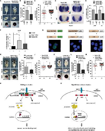
fig.7.a, b, c

Rothe M et al. (2017)
Xla Wt + sipa1l3 MO

NF42 (whole-mount microscopy)
Anatomical Phenotype
abnormal development of eye
abnormal eye morphology
decreased size of the eye
fig.7.h, i, j

Rothe M et al. (2017)
Xla Wt + sipa1l3 MO

NF42 (whole-mount microscopy)
Anatomical Phenotype
abnormal development of eye
abnormal eye morphology
decreased size of the eye