XB-IMG-80819
Xenbase Image ID: 80819
|
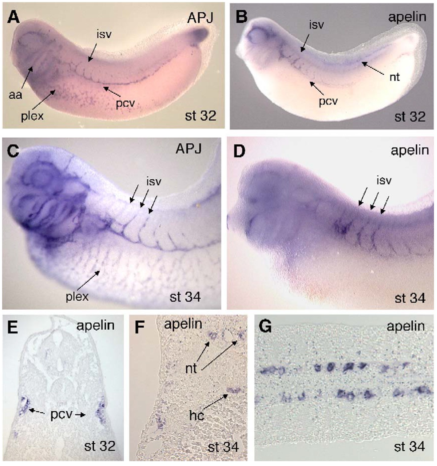
Fig. 2. In situ hybridization analysis of APJ and apelin expression during Xenopus development. (A) In the Xenopus embryo at the early tailbud stage (stage 32) APJ
expression marks developing blood vessels throughout the embryo. The aortic arch region (aa) and the posterior cardinal vein (pcv), ventral vascular plexus (plex) and
branching intersegmental vessels (isv) are indicated. The region of expression in the tailbud does not appear to be associated with vascular tissue. (B) At stage 32,
apelin expression is detected in the anterior region of the posterior cardinal vein, the extending intersegmental vessels and the developing blood vessels surrounding the
eye. Apelin expression is also detected in the ventral neural tube (nt). (C) At stage 35, APJ transcripts are detected throughout the developing vasculature, including the
assembling vascular plexus in ventrolateral regions of the embryo and distinctly in the intersegmental vessels. (D) At stage 35, apelin expression is visible in the
branching intersegmental vessels but expression in the vessels surrounding the eye is now absent. No apelin transcripts are detected in the plexus region. (E) Transverse
section through anterior trunk region of stage 32 embryo stained for apelin transcripts. Note prominent expression in the posterior cardinal veins. (F) Transverse section
through trunk of stage 34 embryo stained for apelin. In addition to posterior cardinal veins, expression is visible in the hypochord (hc) and in paired regions within the
ventral neural tube (nt). (G) Longitudinal section through ventral neural tube of stage 34 embryo stained for apelin. Anterior is to the left. Note that expression is not
continuous but appears to mark paired motor neurons (Saha et al., 1997) (H). Image published in: Cox CM et al. (2006) Copyright © 2006. Image reproduced with permission of the Publisher, Elsevier B. V.
Image source: Published Larger Image Printer Friendly View |