XB-IMG-151901
Xenbase Image ID: 151901
|
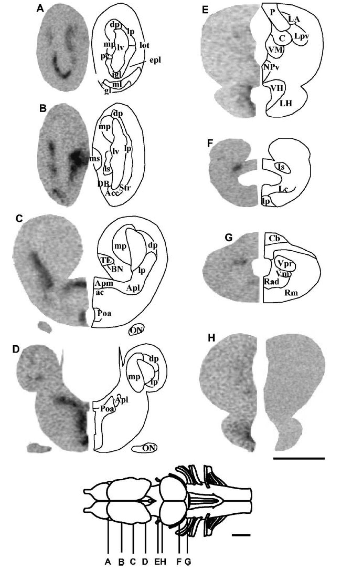
Fig. 3. In situ hybridization histochemistry showing the distribution
of xTRHR1 mRNA in the brain and pituitary of Xenopus laevis.
A–G: Frontal brain sections from white-adapted frogs were hybridized
with an antisense xTRHR1 receptor riboprobe and exposed onto Hyperfilm
max for 2 weeks. H: A control section incubated with a sense
riboprobe (right hemisection) is compared with a consecutive section
hybridized with the antisense probe (left hemisection). See legend to
Figure 1 for other designations. Scale bars 1 mm. Image published in: Bidaud I et al. (2004) Copyright © 2004. Image reproduced with permission of the Publisher.
Image source: Published Larger Image Printer Friendly View |