Eye Regrowth in Xenopus Embryos
Xenopus laevis is a well-established regeneration model, known to regrow spinal cord, tails, larval limbs, and retina. Previously it was thought that embryos had limited ability to repair eye injuries. Kha et al. have found that st. 27 tailbud embryos can readily regrow functional eyes after removal of specified eye tissues. This is a rapid process that happens faster than normal eye development. Following a delay in retinogenesis, regrowing eyes catch up to controls within 3-5 days. The resulting eyes contain the correct eye cell types, connect to the brain, and show visual color preference. Furthermore, this regrowth occurred via the same early mechanism, apoptosis, as found in other regenerative models. Together, these findings demonstrate that frog embryos can successfully repair the eye following tissue loss and that this process requires a common regenerative mechanism. As eye development is well understood in Xenopus, this model will allow for rapid molecular comparisons of successful repair and developmental pathways in the eye.
A model for investigating developmental eye repair in Xenopus laevis.
Exp Eye Res. January 31, 2018; 169 38-47.
Kha CX, Son PH, Lauper J, Tseng KA.
Abstract
Vertebrate eye development is complex and requires early interactions between neuroectoderm and surface ectoderm during embryogenesis. In the African clawed frog, Xenopus laevis, individual eye tissues such as the retina and lens can undergo regeneration. However, it has been reported that removal of either the specified eye field at the neurula stage or the eye during tadpole stage does not induce replacement. Here we describe a model for investigating Xenopus developmental eye repair. We found that tailbud embryos can readily regrow eyes after surgical removal of over 83% of the specified eye and lens tissues. The regrown eye reached a comparable size to the contralateral control by 5 days and overall animal development was normal. It contained the expected complement of eye cell types (including the pigmented epithelium, retina and lens), and is connected to the brain. Our data also demonstrate that apoptosis, an early mechanism that regulates appendage regeneration, is also required for eye regrowth. Treatment with apoptosis inhibitors (M50054 or NS3694) blocked eye regrowth by inhibiting caspase activation. Together, our findings indicate that frog embryos can undergo successful eye repair after considerable tissue loss and reveals a required role for apoptosis in this process. Furthermore, this Xenopus model allows for rapid comparisons of productive eye repair and developmental pathways. It can also facilitate the molecular dissection of signaling mechanisms necessary for initiating repair.
Click here to view this article at Experimental Eye Research.
Click here to view this article on Pubmed.
Click here to view this article on Xenbase.
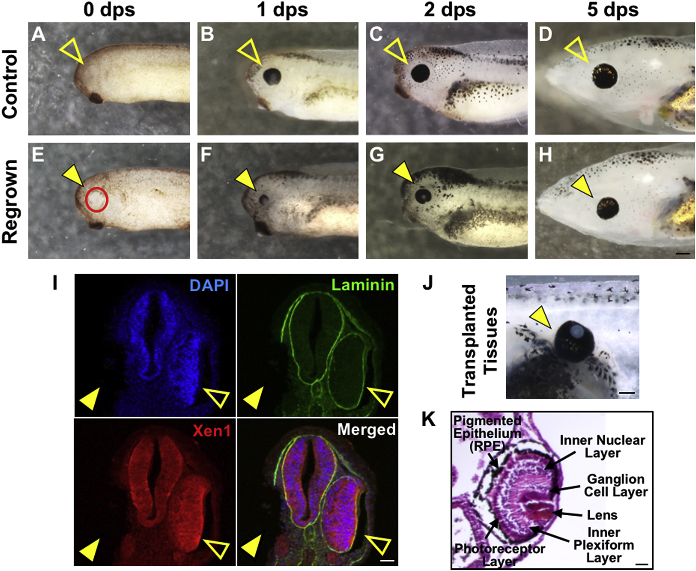
Fig. 1. Tailbud embryos regrow eyes after tissue removal. Images showing normal eye development (A-D) and the eye regrowth process (E-H) starting with st. 27 at the following timepoints: immediately after surgery (0), 1, 2, and 5 days (dps = days post surgery). (A-D) Open arrowheads in the upper panels indicate control, unoperated eyes. (E-H) Closed arrowheads in the bottom panels indicate regrowing eyes. The rate of full eye regrowth (as shown in H and described in Supplemental Fig. 1) was approximately 93% (n = 63). (I) Images are immunostained, transverse sections through the eye plane after surgery. Closed arrowheads indicate surgery site, open arrowheads indicate unoperated eye. Blue color indicates nuclear staining (DAPI). Green color indicates the basal lamina (anti-Laminin), which outlines the optic vesicle. Red color indicates neural tissues (Xen1). (J) Eye tissues removed from a st. 27 embryo were transplanted to its flank at an incision site ∼1/2 from posterior end. By 5 dps, transplanted tissues have a retina and lens similar to the contralateral control eye (not shown) in the same embryo (n > 20). (K) Hematoxylin and eosin stained section of transplanted tissues at 5 dps. (A-H) Up = dorsal, down = ventral, left = anterior, right = posterior. (I) Up = dorsal, down = ventral. Scale bars: A-H = 200 μm, I = 25 μm, J = 200 μm, and K = 50 μm.
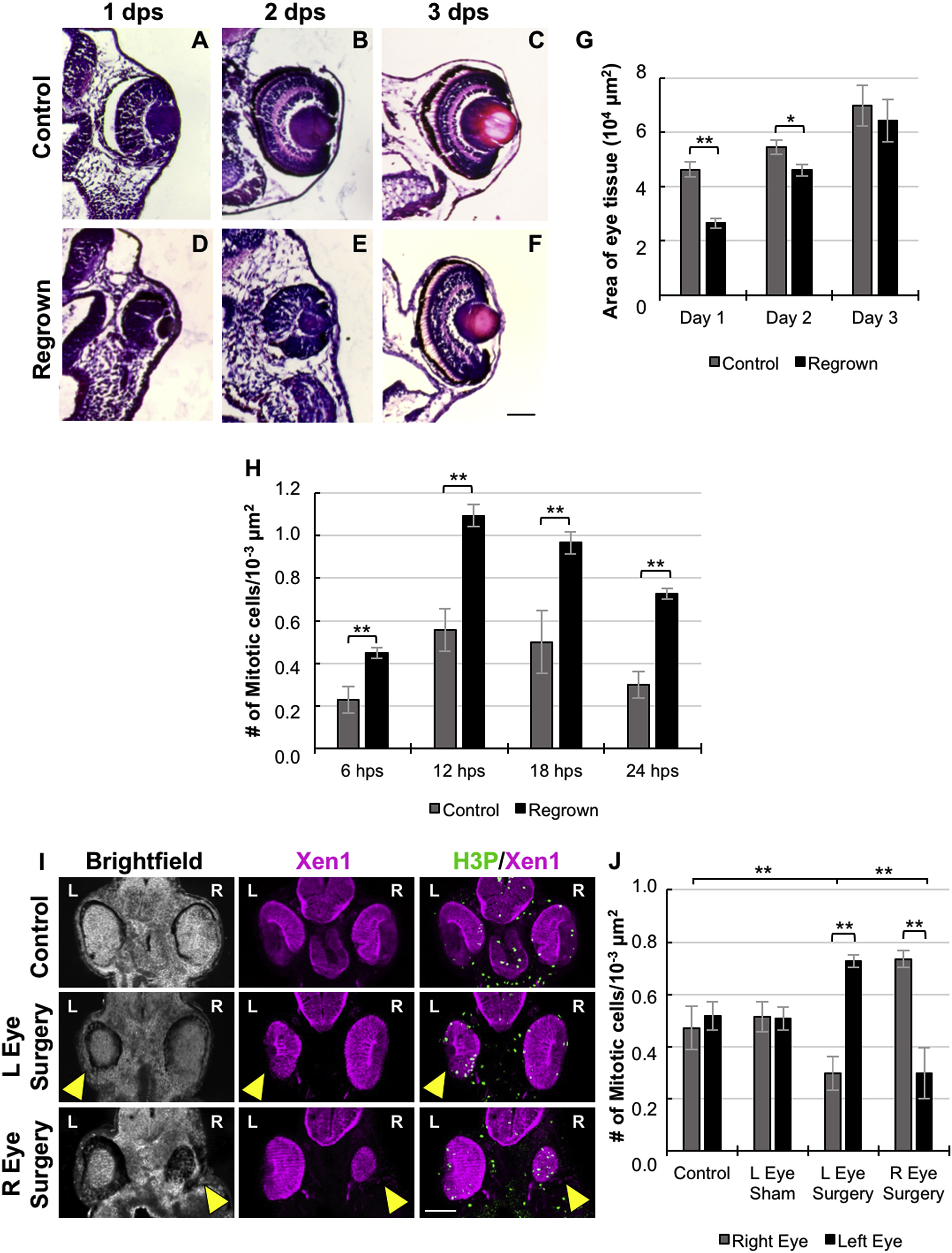
Fig. 2. Proliferation accompanies Xenopus eye regrowth. Hematoxylin and eosin stained sections of unoperated eyes (top panels, A-C) compared to age-matched regrowing eyes (bottom panels, D-F). (D) At 1 dps, a small regrowing eye is seen at surgery site. (E) At 2 dps, retinal layer formation is delayed as compared to control (B). (F) By 3 dps, an eye comparable to control is apparent with similar eye organization and size. Representative images are shown. (G) Timecourse of growth rate after surgery as compared to contralateral unoperated eye at day 1 (57.2%, n = 7), day 2 (84.4%, n = 5), day 3 (92.1%, n = 6). * denotes p < 0.05, ** denotes p < 0.01. Data are means ± SEM. (H) Quantification of mitoses in the regrowing eye structure at the following timepoints: 6, 12, 18, and 24 hps (hps = hours post surgery). There is an increase in the number of mitotic cells in regions where tissue removal surgery was performed compared to the contralateral control side. ** denotes p < 0.01 (n>5 per timepoint). Data are means ± SEM. (I and J) Assessment of mitoses in the regrowing eye at 24 hps. (I) Mitotic cells are indicated in green by anti-phospho Histone H3 signal. Xen1 indicated neural tissues. Closed arrowheads in the brightfield panels indicated regrowing eyes. (J) Quantification of mitoses in embryos under different conditions as shown. Mitoses are increased in regions where tissue removal surgery was performed, but not in the control or sham-operated eye. L indicates left side. R indicates right side. (A-F, I) Up = dorsal, down = ventral. Scale bars: A-F, I = 50 μm.
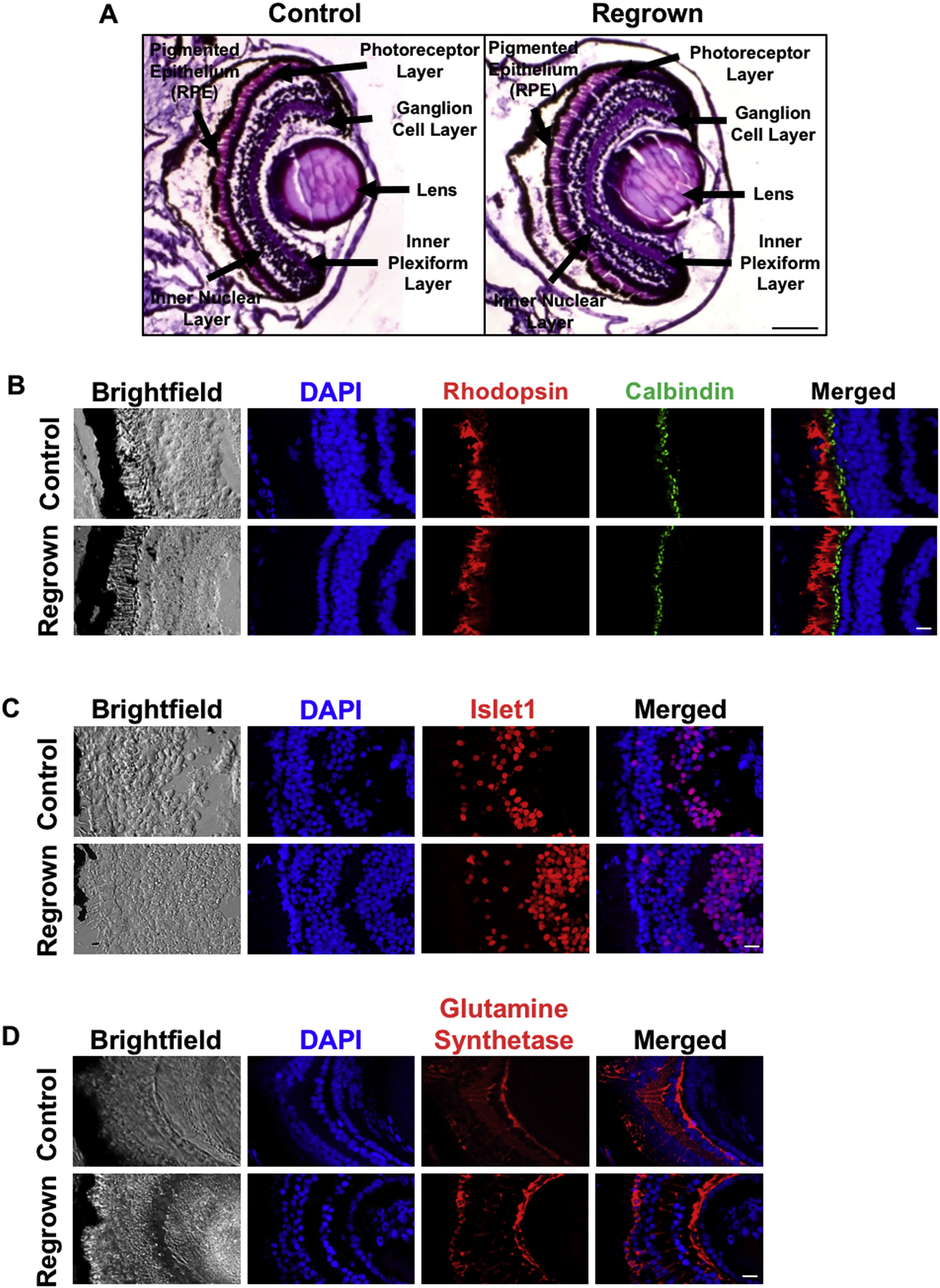
Fig. 3. Regrown eye contains expected cell types. (A) Hematoxylin and eosin stained section of age-matched control (left panel) and regrown eyes at 5 dps (right panel). Regrown eye contains same eye structures compared to unoperated sibling control. (B-D) Identification of retinal cell types in eye regenerates. Dark pigmented tissue surrounding the retina is the retinal pigmented epithelium (RPE). (B) Eye sections stained with DAPI (DNA), anti-Rhodopsin antibody (rod photoreceptors), and anti-Calbindin-D-28K antibody (cone photoreceptors). (C) Eye sections stained with DAPI, anti-Isl1 antibody (identifies retinal ganglion cells, amacrine cells, bipolar cells, and horizontal cells). (D) Eye sections stained with DAPI, and anti-Glutamine Synthetase (Müller glia). (A-D) Up = dorsal, down = ventral. Scale bars: A-D = 50 μm.
Adapted with permission from Elsevier: Kha et al. (2018). A model for investigating developmental eye repair in Xenopus laevis. Exp Eye Res. January 31, 2018; 169 38-47. Copyright 2018.
This work is licensed under a Creative Commons Attribution 4.0 International License. The images or other third party material in this article are included in the article’s Creative Commons license, unless indicated otherwise in the credit line; if the material is not included under the Creative Commons license, users will need to obtain permission from the license holder to reproduce the material. To view a copy of this license, visit http://creativecommons.org/licenses/by/4.0/