XB-IMG-158390
Xenbase Image ID: 158390
|
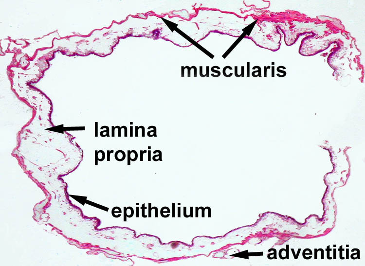
Low magnification image of the urinary bladder. The lumen of the bladder is lined with transitional epithelium. Underlying the epithelium is a loose connective tissue called the lamina propria. The next layer is a layer of smooth muscle cells called the muscular. The outer layer is either an adventitia or serosa.
Image from AF Wiechmann and CR Wirsig (2003) "Color Atlas of Xenopus laevis Histology", (page 57, Chapter 6,Urinary system: Figure 1). Copyright 2003. Kluwer Academic Publishers. Reproduced with kind permission from Springer Science & Business Media B.V.
Image from AF Wiechmann and CR Wirsig (2003) "Color Atlas of Xenopus laevis Histology", (page 66, Chapter 6,Urinary system: Figure 20). Copyright 2003. Kluwer Academic Publishers. Reproduced with kind permission from Springer Science & Business Media B.V. Image published in: Color Atlas of Xenopus laevis Histology Larger Image Printer Friendly View |