XB-IMG-158229
Xenbase Image ID: 158229
|
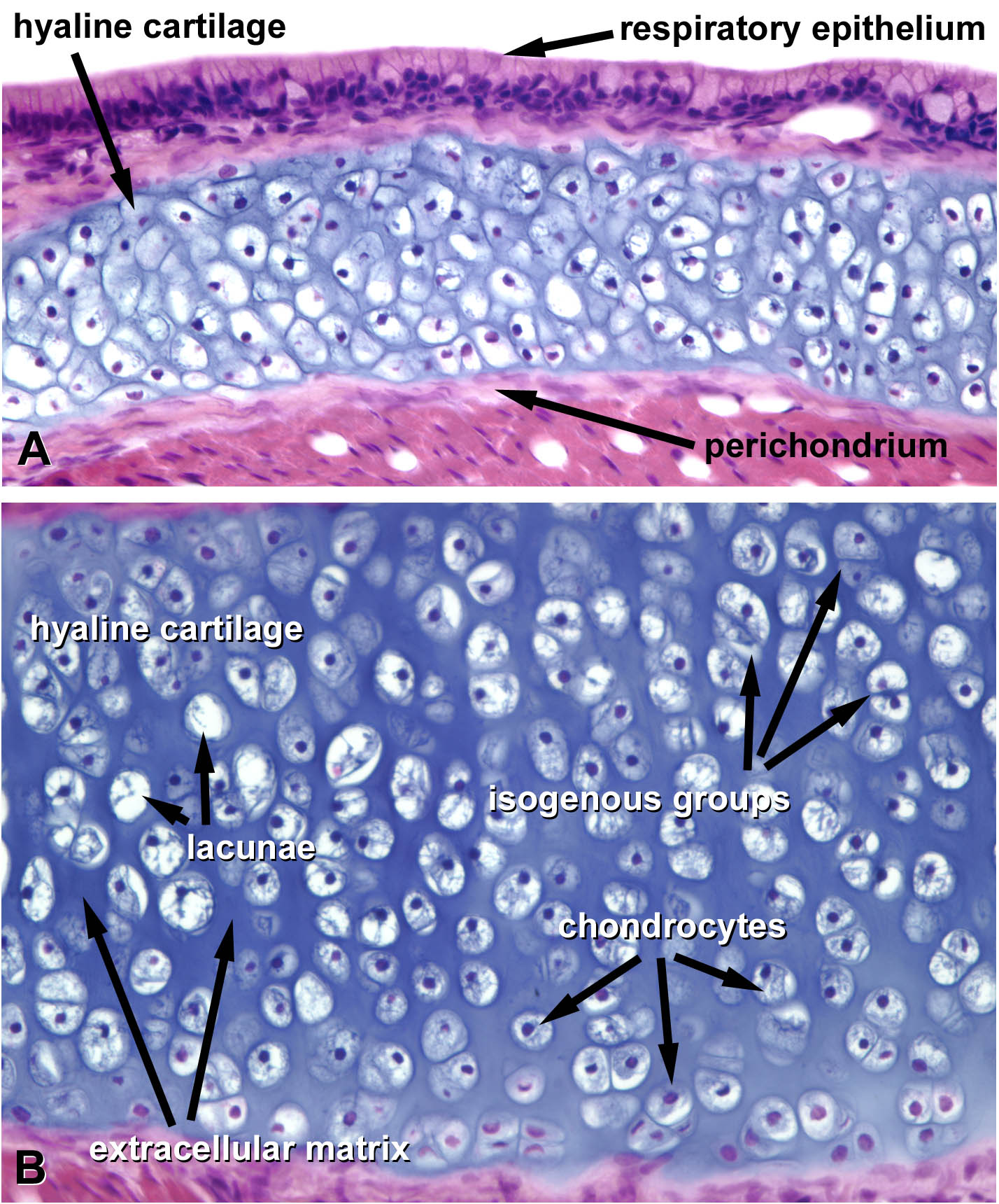
A. Low magnification image of the hyaline cartilage that provides structural support for the trachea. Note that the cartilage is completely surrounded by a dense connective tissue perichondrium, which is one source of new chondrocytes. B. High magnification of the trachea showing lacunar spaces occupied by chondrocytes. Chondrocytes are not well-preserved during fixation, but the nuclei can be discerned. Isogenic groups are clusters of daughter chondrocytes that have dived, and will produce additional basophilic matrix resulting in interstitial growth.
Image from AF Wiechmann and CR Wirsig (2003) "Color Atlas of Xenopus laevis Histology", (page 9, Chapter 1, Basic tissues: Figure 15). Copyright 2003. Kluwer Academic Publishers. Reproduced with kind permission from Springer Science & Business Media B.V.
Image published in: Color Atlas of Xenopus laevis Histology Larger Image Printer Friendly View |